

Anti-I type diabetes fuse protein and preparation thereof
A fusion protein, fusion gene technology, applied in the directions of botanical equipment and methods, biochemical equipment and methods, peptide/protein components, etc., to achieve the effect of relieving pain and the threat of infection and stabilizing gene expression products
- Summary
- Abstract
- Description
- Claims
- Application Information
AI Technical Summary
Problems solved by technology
Method used
Image

Examples
Embodiment 1
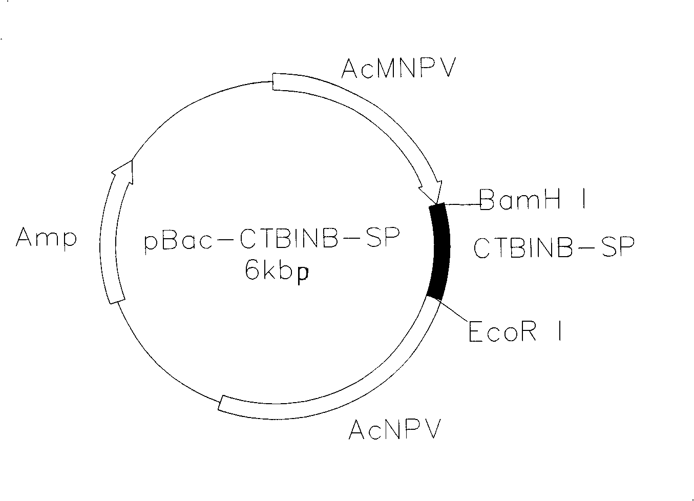
[0029] (1) Construct the transfer plasmid pBac-CTBINB-SP containing the fusion gene of CTB and human insulin B chain antigen epitope.
[0030] Two primers P1 and P2 of the sequences shown in SEQ ID No: 2 and SEQ ID No: 3 were designed.
[0031] With the pBac-CTBINS plasmid (seeing No. 200310121132.1 patent application) as a template, the sequences shown in SEQ ID No: 2 and SEQ ID No: 3 are used as primers to amplify to obtain a fusion gene CTBINB-SP with a size of about 430bp, which is obtained by PCR The target fragment was digested by BamHI and EcoRI, and connected to the transfer vector pBacPAK8, which was also digested by BamHI and EcoRI, to construct the bacmid transfer plasmid pBac-CTBINB containing the fusion gene of CTB and human insulin B chain antigen epitope -sp.
[0032] (2) Obtaining the recombinant silkworm baculovirus BmBacCTBINB-SP containing the fusion gene of CTB and human insulin B chain antigen epitope: the transfer plasmid pBac-CTBINB-SP was co-transfecte...
experiment example 1
[0035] [Experimental example 1] Construction of transfer plasmid pBac-CTBINB-SP containing fusion gene
[0036] The pBac-CTBINS plasmid (see my patent application (patent number: 200310121132.1)) was used as a template and P1 and P2 were used as primers for amplification. The reaction conditions were pre-denaturation at 94°C for 5 minutes, amplification at 94°C for 1 minute, and 57°C 1 minute, 1 minute at 72°C, react for 30 cycles, and finally incubate at 72°C for 10 minutes to obtain the target fusion gene CTBINB-SP with a size of about 430bp, and the 5' and 3' ends of the gene have BamHI and EcoRI respectively point. The above target fusion gene was digested with BamHI and EcoRI and then connected to pBacPAK8 (CLONTECH Company) which was also digested with BamHI and EcoRI to construct the baculovirus transfer plasmid pBac- For CTBINB-SP, after the correct insertion of the gene was identified by enzyme digestion analysis and PCR, automatic sequence determination showed that ...
experiment example 2
[0037] [Experimental example 2] Obtaining recombinant baculovirus of CTB and human insulin B chain antigen epitope fusion gene
[0038] Take 5ul insect baculovirus transfer plasmid pBac-CTBINB-SP containing the fusion gene of CTB and human insulin B chain antigen epitope and 6ul wild silkworm nuclear polyhedrosis virus DNA for co-transfection. Take 6ul Lipofectin (GIBCOBRL company) and add 100ul serum-free TC-100 medium and mix well. The BmN cells previously cultured in a 35mm Dish were washed twice with serum-free TC-100 (GIBCOBRL company) medium, and the transfer plasmid and Lipofectin mixture was added dropwise, cultured at 27°C for 4-5 days, and the supernatant was collected for the second stage. One round of plaque screening. Take 5ul of the supernatant to infect the BmN cells in a 35mm Dish, discard the supernatant after 1 hour and add an equal amount of mixed TC-100 medium and low melting point agarose. Pick plaques after 4-5 days, infect BmN cells for 3-4 days, save ...
PUM
 Abstract
Description
Claims
Application Information