
Special shape tissue engineering bracket and construction method and use thereof

A tissue engineering scaffold and a technology with a specific shape, which are applied in the field of tissue engineered cartilage preparation, can solve problems such as being unfavorable for industrialized production, complex modeling of an all-external nose model stent, and unfavorable for the construction of bone marrow stromal stem cells, etc.

Inactive Publication Date: 2009-02-11
SHANGHAI TISSUE ENG LIFE SCI
View PDF0 Cites 25 Cited by
  • Summary
  • Abstract
  • Description
  • Claims
  • Application Information

AI Technical Summary

Problems solved by technology

However, it has always been a big problem to process and pre-shape the cotton-like non-woven PGA fibers into precise shapes of auricles and external noses.
[0004] The auricle model brackets made in the existing reports are all obtained by manual modeling of healthy auricles, and the obtained auricles are autologous normal auricles, which lack clinical application value
At the same time, it has been reported that gel scaffolds are often used for direct implantation in vivo, which is not conducive to the use of human body, nor is it conducive to the use of bone marrow stromal stem cells as seed cells for construction, and it is not conducive to industrial production.
There is no report on the full external nose model bracket due to the complexity of model taking

Method used


Image

  • Special shape tissue engineering bracket and construction method and use thereof
  • Special shape tissue engineering bracket and construction method and use thereof
  • Special shape tissue engineering bracket and construction method and use thereof

Examples


Embodiment 1

[0094] Fabrication of personalized auricle prosthetic bracket

[0095] Materials and methods

[0096] 1. Materials

[0097] 1.1 Equipment

[0098] 16-slice spiral CT (GE LIGHT SPEED 16 SLICES) purchased from General Corporation; medical tomography processing software purchased from Shanghai Jiaotong University; rapid prototyping machine purchased from Z CORPORATION.

[0099] 1.2 Clinical medical records

[0100] Randomly selected 3 clinical cases of microtia from the Department of Plastic and Reconstructive Surgery of the Ninth People's Hospital Affiliated to Shanghai Jiao Tong University School of Medicine.

[0101] 2. Method

[0102] 2.1 CT scan

[0103] The whole head of case A was scanned with continuous thin-slice CT volume. The scanning equipment is a 16-slice spiral CT of the American General Company, each slice is 0.625mm, and an average of 136 slices. The rotation angle is 216°, and the exposure time is 0.1S. All the images are transmitted to the graphics work...

Embodiment 2

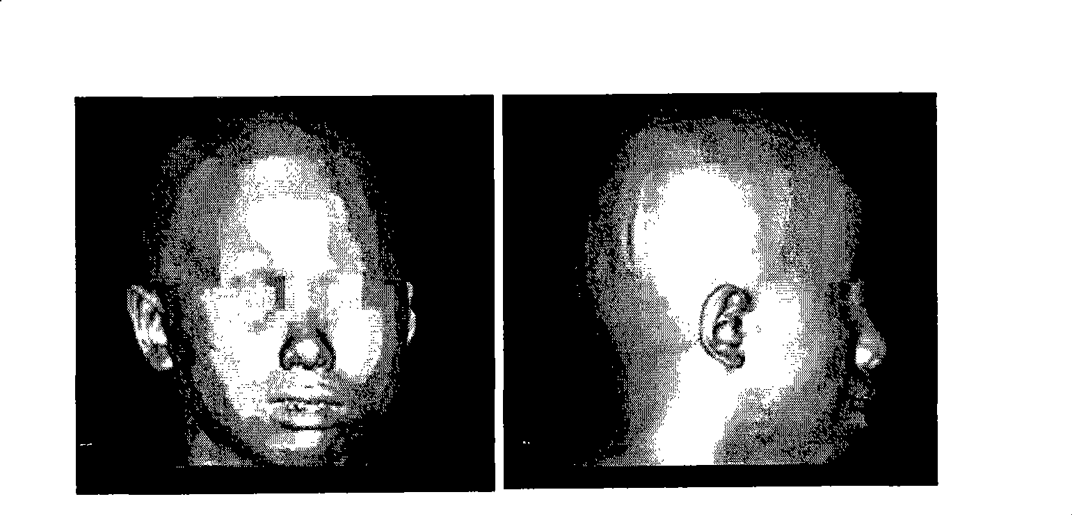
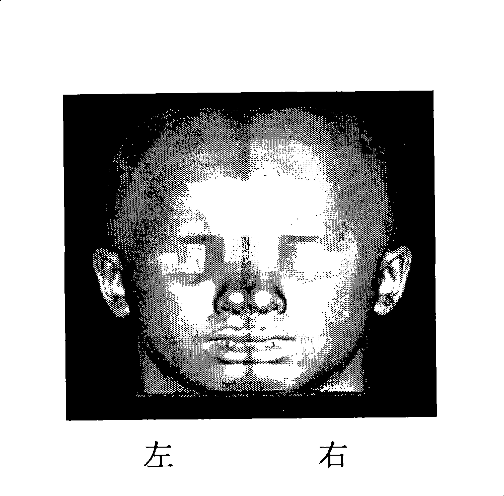
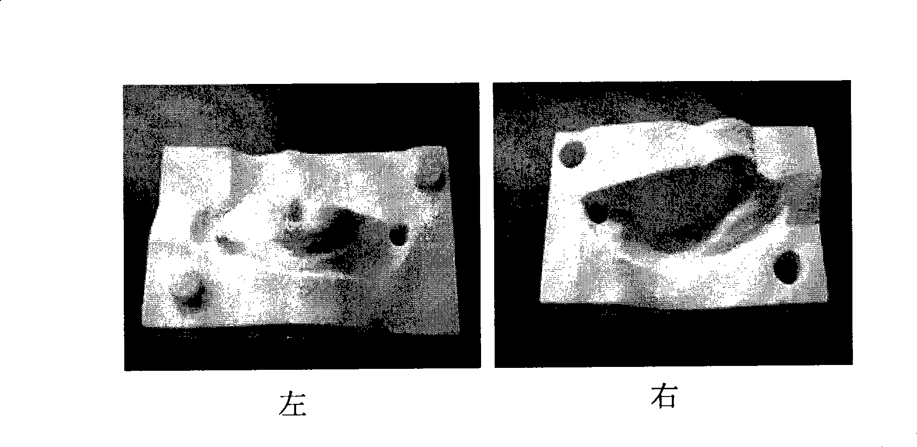
[0122] Fabrication of personalized external nasal restorations

[0123] Materials and methods

[0124] 1. Materials

[0125] 1.1 Equipment

[0126] 16-slice spiral CT (GE LIGHT SPEED 16 SLICES) purchased from General Corporation; medical tomography processing software purchased from Shanghai Jiaotong University; rapid prototyping machine (SpectrumZ510) purchased from Z CORPORATION.

[0127] 1.2 Clinical medical records

[0128] Randomly selected 3 clinical cases of plastic surgery in Ninth People's Hospital Affiliated to Shanghai Jiaotong University School of Medicine.

[0129] 2. Method

[0130] 2.1 CT scan

[0131] In this case, serial thin-slice CT volume scans were performed on the whole head. The scanning equipment is a 16-slice spiral CT of the American General Company, each slice is 0.625mm, and an average of 136 slices. The rotation angle is 216°, and the exposure time is 0.1S. All the images are transmitted to the graphics workstation of GE Company through the...

Embodiment 3

[0148] Fabrication of cartilage for auricle restoration

[0149] Carry out electron microscope scanning to the auricle prosthesis bracket that embodiment 1 makes, determine optimal PLA: (PGA+PLA) ratio ( Figure 8 , 9 ), the result is preferably 20%-30%. Composite the porcine chondrocytes with the above-mentioned auricle prosthetic scaffold, wherein the concentration of porcine chondrocytes in the complex is 5×10 7 cells / cm 3 . They were cultured in vitro, and samples were collected at 4 weeks, 8 weeks and 12 weeks respectively. Histological evaluation (mass morphology, HE staining, Saffranin'0 staining and type II collagen immunohistochemical staining) was performed. See Figure 10 .

[0150] The results show that auricular grafts with individualized shapes can be obtained.

[0151] discuss

[0152] Chondrocytes are the earliest and most commonly used seed cells in tissue engineering cartilage research. However, obtaining cells from the body's own cartilage tissue wil...


PUM


Abstract

The invention discloses a method for in vitro establishment of special-shaped tissue engineering scaffolds, comprising steps as follows: (1) applying computer aided design to establish a digital model for special-shaped tissues or organs; (2) guiding the digital model acquired in the step (1) into a rapid prototyping machine for processing, to shape a solid female die for the special-shaped tissues; (3) adding biodegradable materials acceptable by the medical science into the solid female die gained in the step (2), to acquire a special-shaped tissue engineering scaffold. The special shapes are chosen according to the shapes of auricle, external nose, nose prosthesis, chin prosthesis and other human issues or organs. The special-shaped tissue engineering scaffold can be effectively compounded with the chondrocytes to finally form special-shaped tissue engineering cartilages. The invention also provides a detailed method of preparing tissue engineering auricle and external nose scaffolds and application thereof.

Description

technical field [0001] The invention relates to the fields of medicine and biomedical engineering, and more particularly to a preparation method and application of tissue-engineered cartilage applied in the field of craniomaxillofacial surgery of plastic surgery. Background technique [0002] The defect or loss of the outer ear caused by congenital microtia, trauma, or tumor is relatively common in clinical practice; the current treatment method is mainly to excise the autologous costal cartilage, and sculpt it to reconstruct the affected ear; however, there are different shapes in different degrees. , secondary defects and other issues. The defect or loss of the external nose due to congenital external nasal deformities, trauma, and tumors is relatively common in clinical practice; the current treatment methods are mainly the use of frontal pedicled flaps and postauricular island flaps with superficial temporal postauricular vascular pedicles. , forearm leather tube, and m...

Claims


Application Information

Patent Timeline
Patent Type & Authority Applications(China)
IPC IPC(8): A61F2/00G06F17/50A61B6/03G06T1/20G06T15/00A61L27/14A61L27/50A61L31/04
Inventor 周广东曹谊林张路刘伟崔磊
Owner SHANGHAI TISSUE ENG LIFE SCI
Learn More