Single-optical fiber multiphoton fluorescence scanning endoscope

A multi-photon, endoscope technology, applied in the field of optical systems, can solve the problems of mechanical inertia, image motion blur, unfavorable scanning speed, etc., and achieve the effect of simple driving, convenient use, and overcoming mechanical inertia.

Inactive Publication Date: 2009-07-22
ZHEJIANG GONGSHANG UNIVERSITY
View PDF1 Cites 29 Cited by
  • Summary
  • Abstract
  • Description
  • Claims
  • Application Information

AI Technical Summary

Problems solved by technology

The multi-photon scanning mechanisms and methods recorded in the current literature and patents all use micromechanical mechanisms, or piezoelectric crystals to drive the end of the optical fiber, and scan the target area along a certain horizontal and vertical axes. Since the mechanical vibration is nonlinear vibration, and The structure is complex, the production cost is high, and there is mechanical inertia, which is not conducive to the improvement of scanning speed, and the human organs are always wriggling, resulting in motion blur of the image

Method used


Image

  • Single-optical fiber multiphoton fluorescence scanning endoscope
  • Single-optical fiber multiphoton fluorescence scanning endoscope

Examples


Embodiment Construction

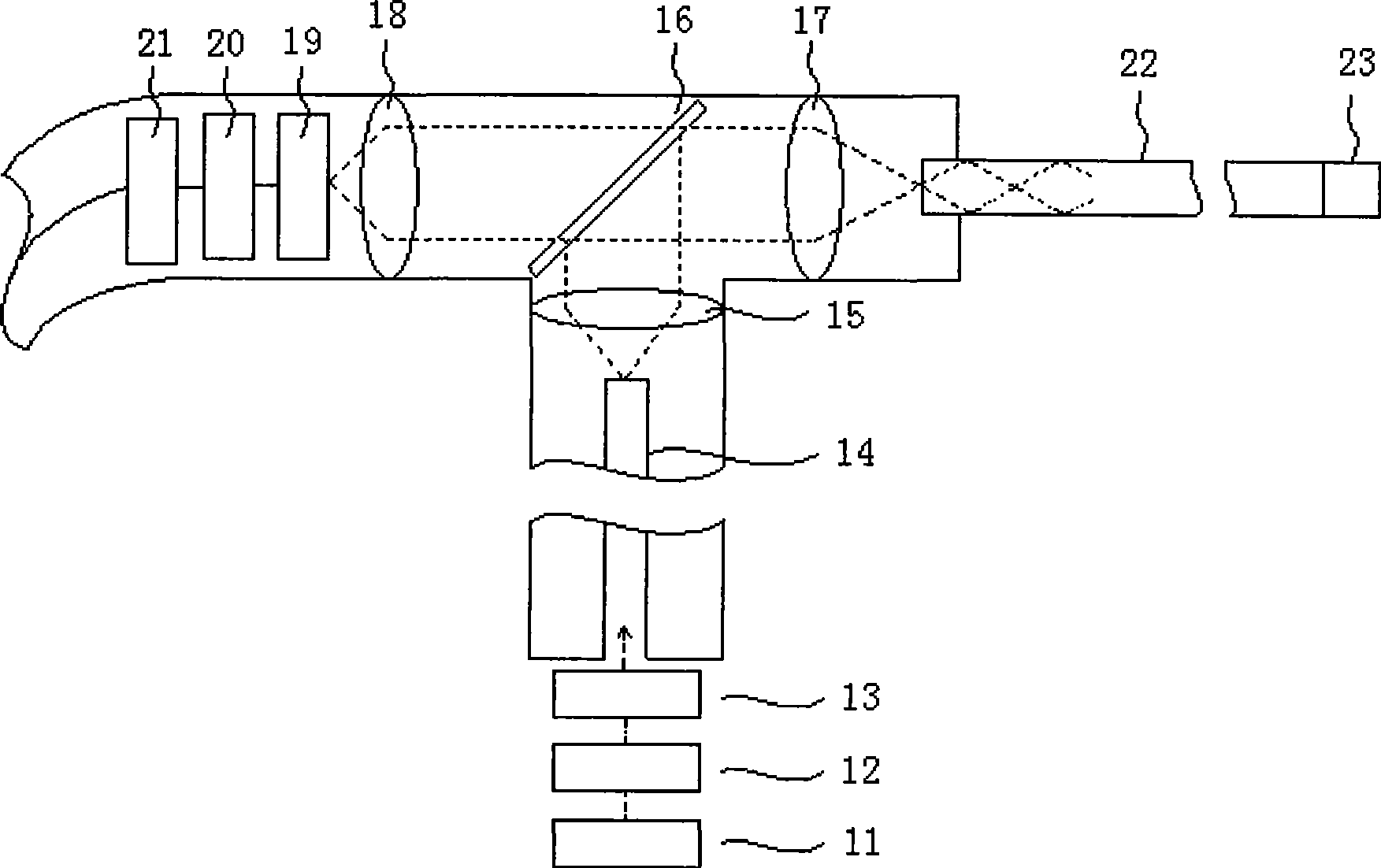
[0022] like figure 1 The system optical path diagram of the shown single-fiber multiphoton fluorescence scanning endoscope, the present invention adopts titanium sapphire laser 11 as the excitation light source, the output center wavelength is 800nm, the laser of bandwidth 10nm; Through electro-optic modulator 12, produce ultrashort pulse laser; Since the laser beam passes through the acousto-optic deflector to generate temporal and spatial dispersion, a dispersion compensation unit 13 is installed at the incident end of the laser; the generated excitation laser passes through a single single-mode optical fiber 14, a lens 15, is reflected by a beam splitter 16, and passes through After the lens 17 is focused, it is coupled into a single single-mode optical fiber 22, which is carried in the endoscope duct and enters the body. The front end of the single-mode optical fiber 22 is an acousto-optic deflection scanning front end 23 .

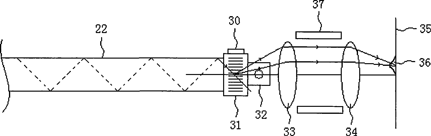
[0023] like figure 2 The structure diagram o...


PUM


Abstract

The invention relates to a single-fiber multi-photon fluorescent scanning endoscope, applied in early screening and diagnosis of various mucosa cancers of human body. A single mode fiber is carried inside an endoscope system to enter a human body and guide extracorporeal ultrashort pulse laser into intracorporeal target cell, and an acousto-optic deflector is adopted to carry out laser deflexion in two orthogonal directions; moreover, the endoscope adopts a piezocrystal to perform fine adjustment on the focus of the lens group so as to adjust fluorescence excitation depth, and adopts a multi-photon fluorescent imaging mechanism to eliminate confocal pinholes; therefore, the endoscope reduces the process difficulty of making acousto-optic deflexion scanning front end and also overcomes mechanical inertia of devices adopting micromechanics to realize deflexion scanning so as to ensure convenient integrated molding and miniaturization of an entire scanning imaging tail end. The system provided by the invention can carry out cell morphologic analysis on suspect cancer cells at low cost during routine endoscope examination.

Description

technical field [0001] The invention relates to an optical system, in particular to an optical system applied in the technical field of early screening and diagnosis of various mucosal cancers of the human body. Background technique [0002] The current gold standard for cancer diagnosis is the pathological section of diseased tissue cells. Diagnostic doctors use a microscope to zoom in on the pathological section to obtain the morphological characteristics of the tissue cells, so as to determine whether the pathological section contains cancerous cells. [0003] Although the method of pathological section is of great significance for the diagnosis of advanced cancer, but this method is invasive, the diagnosis cycle is long, and it may cause the artificial spread of cancer cells in the process of obtaining tissue cells; on the other hand, early detection of cancer can Greatly improve the cure rate of cancer and prolong the survival period of cancer patients. [0004] For so...

Claims


Application Information

Patent Timeline
Patent Type & Authority Applications(China)
IPC IPC(8): A61B1/07
CPCA61B5/0071A61B5/0084
Inventor 凌云邢建国朱安定赵文敏
Owner ZHEJIANG GONGSHANG UNIVERSITY
Why Eureka
Try Eureka