A group of antigen epitope polypeptide and uses thereof

A technology of antigenic epitopes and polypeptide chains, applied in the field of molecular immunology, can solve the problem of small differences in antigenicity

Inactive Publication Date: 2009-07-29
HARBIN VETERINARY RES INST CHINESE ACADEMY OF AGRI SCI
View PDF0 Cites 15 Cited by
  • Summary
  • Abstract
  • Description
  • Claims
  • Application Information

AI Technical Summary

Problems solved by technology

[0009] Although the various strains of Japanese encephalitis virus often have obvious differences in virulence an

Method used


Image

  • A group of antigen epitope polypeptide and uses thereof
  • A group of antigen epitope polypeptide and uses thereof
  • A group of antigen epitope polypeptide and uses thereof

Examples


Embodiment 1

[0039] Example 1 Overlapping polypeptide fusion expression series of JEV E protein antigen polypeptide fragments and indirect ELISA screening of JEV E protein B cell antigen epitopes

[0040] According to the amino acid sequence of the E protein of JEV (SA14-14-2 strain), a series of 50 peptides in total were designed. These polypeptides overlap each other and cover the region of 1-402 amino acid residues of the JEV E protein, which contains the E protein domain. I (Domain I) and domain II (Domain II). According to the amino acid sequence of each polypeptide, a pair of DNA chains are designed and synthesized, and the DNA chains are inserted into the expression vector pGEX-6p-1 and GST for fusion expression. The series of recombinant proteins expressed in fusion and anti-JEV serum were analyzed by indirect ELISA to analyze the epitope of JEV E protein. The results are shown in Table 1.

[0041] The specific process is as follows:

[0042] Design the polypeptide sequence→synthesize ...

Embodiment 2

[0049] Example 2 Location of core sequence of antigen epitope

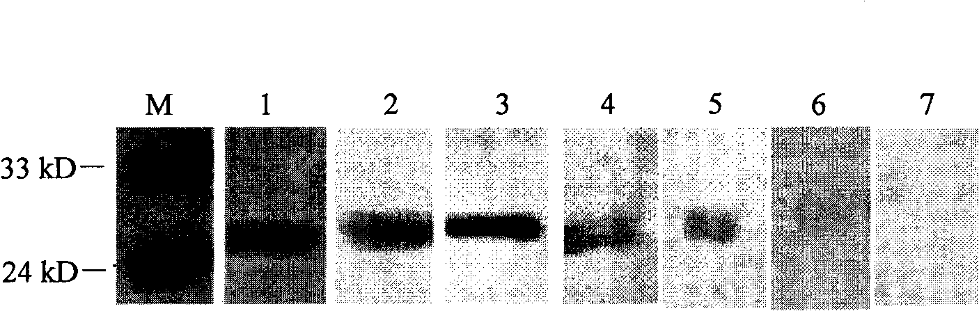
[0050] According to the preliminarily identified epitope sequence in Example 1, further design small fragment polypeptides, or design a series of polypeptides that are reduced by one amino acid residue from C-terminus to N-terminus and those that are reduced by one amino acid residue sequentially from N-terminus to C-terminus. A series of peptides were fused and expressed with JEV positive serum for Western blot analysis. The two ends were reduced to the point that the peptide fusion protein lost the ability to bind to JEV positive serum to determine the core sequence of the epitope. The reaction results are shown in Table 2.

[0051] Table 2 Indirect Western blot reaction results of peptide fusion protein and anti-JEV serum

[0052] serial number

Codename

GST fusion peptide sequence

Western blot test results

SEQ ID NO: 1

E1

FNCLGMGNRDFIEGAS

+++

SEQ ID NO: 51

E...

Embodiment 3

[0058] Example 3 Mutation analysis of epitope core sequence

[0059] The epitope core sequence determined in Example 2 was compared with the E protein sequence of different strains of Japanese encephalitis and compared with the Meruo Valley Encephalitis Virus (MVEV), West Nile Virus (WNV), and Usutu Virus (Usutu Virrus, UV), Kunjin virus (Kunjin virus, KUN) and Dengue virus (Dengue Virus, DV) and other flavivirus E proteins were analyzed and compared by homology.

[0060] To compare the homology of the epitope core sequence determined in Example 2 with other viruses, first select short peptides of homologous sequences with a homology greater than 50%, which are SEQ ID NO: 64, SEQ ID NO: 95, SEQ ID NO :108 and SEQ ID NO: 124, after GST fusion expression, Western blot analysis of the reactivity with JEV positive serum, the reaction results are shown in Table 3.

[0061] Table 3 Western blot reaction results of peptide fusion protein and anti-JEV serum

[0062]

[0063]

[0064] ...


PUM


Abstract

The invention discloses a set of epitope polypeptides, particularly relates to a neutralizing B cell epitope sequence of encephalitis B virus E protein, and further discloses application of controlling and diagnosing encephalitis B virus by these epitopes. The amino acid sequences of the epitope polypeptides in the invention are respectively any amino acid sequence of SEQ ID NO: 64, SEQ ID NO: 79, SEQ ID NO: 95, SEQ ID NO: 108, SEQ ID NO: 124 and SEQ ID NO: 139. After the epitope polypeptides in the invention are coupled or fused with carrier proteins to be expressed as immunogenic or vaccine immuno animal organism, neutralizing antibodies aiming at JEV can be generated, and the JEV can be neutralized in vivo or in vitro so as to prevent virus from infecting animal organism. The epitope polypeptides in the invention or the junctional complex thereof can be used as reagents for detecting encephalitis B virus antibodies or encephalitis B virus polypeptide antibodies.

Description

Technical field [0001] The present invention relates to epitope polypeptides, in particular to Japanese encephalitis virus E protein neutralizing B cell epitope polypeptides. The present invention also relates to the application of the epitope polypeptides in the prevention and diagnosis of Japanese encephalitis virus. The field of molecular immunology. Background technique [0002] Japanese encephalitis Japanese encephalitis is an important mosquito-borne zoonotic disease caused by Japanese Encephalitis virus (JEV). The virus was first isolated from the brain tissue of patients in Japan in 1953, so it is called Japanese encephalitis virus, and the disease caused by it is called Japanese encephalitis in Japan. This virus was first isolated in my country in 1940. In order to distinguish it from type A encephalitis, it was named Japanese encephalitis, or Japanese encephalitis for short. There are 30,000-50,000 cases of Japanese encephalitis worldwide each year, about 25%-30% die, a...

Claims


Application Information

Patent Timeline
IPC IPC(8): C07K7/06C12N15/40A61K39/12A61P31/14G01N33/68C12R1/93
CPCY02A50/30
Inventor 华荣虹童光志步志高
Owner HARBIN VETERINARY RES INST CHINESE ACADEMY OF AGRI SCI
Why Eureka
Try Eureka