Method for cutting Yersinia pestis F1 antigen and relative application thereof

A technology of F1 antigen and Yersinia pestis, which is applied in the field of analyzing the reactivity between protein A antibody-positive samples and protein A linear epitope, can solve the problems of unreasonable step distance, omission, and limited application

Inactive Publication Date: 2009-09-16
ICDC CHINA CDC
View PDF0 Cites 3 Cited by
  • Summary
  • Abstract
  • Description
  • Claims
  • Application Information

AI Technical Summary

Problems solved by technology

However, due to the limitations of current technology, for example, in the current research methods for determining which site or sites of the antigen molecule the antibody in the sample is targeting, the method of chemically synthesizing polypeptides and cloning and expressing polypeptides is a relatively common method, but these The method often requires an overlapping step-by-step design for the full length of the protein, and the workload is relatively large, and often due to unreasonable step distances, some sites are missed or even no reactive sites can be found; proteolysis or cutting proteins and Combining fragment separation technology, mass spectrometry identification and other technologies to find and determine reactive sites is also a method for analyzing epitopes, but this method is often difficult to determine the specific cleavage scheme and find a suitable cleavage reagent after analyzing the composition of the protein itself. As a result, its application is limited. At present, there is no progress in using substantive methods to explain and verify the positive serum F1 antibody in natural populations.

Method used


Image

  • Method for cutting Yersinia pestis F1 antigen and relative application thereof
  • Method for cutting Yersinia pestis F1 antigen and relative application thereof
  • Method for cutting Yersinia pestis F1 antigen and relative application thereof

Examples


Embodiment 1

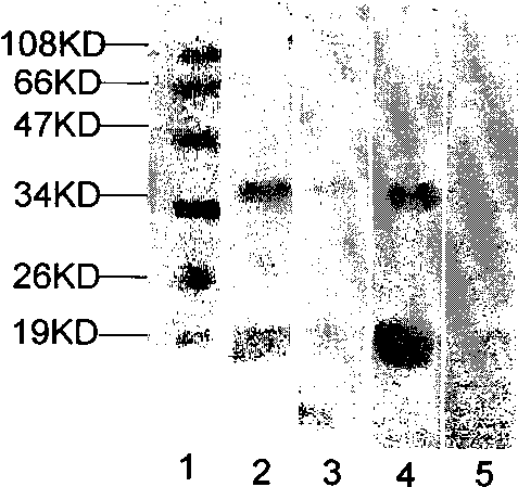
[0055] Embodiment 1, the cleavage of F1 antigen

[0056] In this embodiment, hydroxylamine (Hydroxylamine) is mainly used as a cutting agent to cut the F1 antigen so that the linear epitopes belong to different cutting fragments.

[0057] 1. Experimental materials:

[0058] 1. F1 antigen: purchased from Qinghai Provincial Center for Disease Control and Prevention, endemic disease prevention and control, natural F1 antigen;

[0059] 2. Hydroxylamine: Fluka reagent, ~50% in H 2 O, (RT);

[0060] 3.1.5M Tris-Cl (pH9.0): Weigh 9.06 g of Tris, add 48.5 ml of double distilled water, add about 1.5 ml of 37% concentrated HCl, and adjust the pH to 9.0.

[0061]2. Experimental method:

[0062] 1. Weigh 1 mg of F1 antigen and dissolve it in 50 μl of 1.5M Tris-Cl (pH9.0);

[0063] 2. Add 50 μl of 50% hydroxylamine to the above solution containing F1 antigen, mix gently with a pipette, and be careful not to generate air bubbles;

[0064] 3. Transfer the above mixed solution into a me...

Embodiment 2

[0069] Example 2, Renaturation of F1 antigen cleavage product

[0070] This embodiment mainly removes the chemical cutting agent hydroxylamine in the cleavage product obtained in Example 1, and refolds the cleavage product. The main operations are as follows:

[0071] 1. Reagents and instruments

[0072] 1. Concentrated HCl: effective content 36% to 38%, Beijing Chemical Factory;

[0073] 2. F1 cleavage product: the product obtained in Example 1;

[0074] 3. Instruments: freeze dryer (Christ); ultra-low temperature refrigerator (-80°C);

[0075] 2. Experimental method

[0076] 1. Dispense the F1 cleavage product in 10 μl / tube;

[0077] 2. Add 100 μl of double distilled water to each tube and mix gently;

[0078] 3. Put each aliquot into a -80°C refrigerator and freeze for about 2 hours;

[0079] 4. Quickly transfer to a freeze dryer (pre-cooling for 20 minutes), freeze-drying, the temperature of the cold well is below -50°C, and the vacuum degree reaches ≤0.07mbar, and ...

Embodiment 3

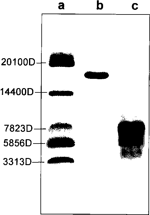
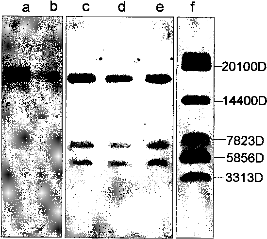
[0083] Example 3, electrophoresis separation of F1 antigen cleaved fragments

[0084] In this example, Tricine-SDS-PAGE is used for small molecule electrophoresis. The gel is divided into separating gel, spacer gel and stacking gel, which are 7cm, 2cm, and 1cm respectively. The electrophoresis conditions are 60V1h, 80V4h, high ionic strength in the sample, and about 2μl The amount of sample loaded ensures electrophoretic resolution.

[0085] 1. Equipment and reagents

[0086] 1. Equipment: electrophoresis apparatus (amersham pharmacia biotech, Hoefer miniVE), image scanner (amersham pharmacia biotech, ImageScanner), decolorization shaker.

[0087] 2. Electrophoresis buffer:

[0088] Negative electrode buffer: Tricine (Solarbio): 17.92g

[0089] Tris: 12.114g

[0090] SDS: 1g

[0091] h 2 O dilute to 1000ml

[0092] Positive electrode buffer: Tris: 24.228g

[0093] Concentrated HCl: 1.5ml

[0094] h 2 O dilute to 1000ml

[0095] 3. The preparation of 49.5% (0.495T) g...


PUM


Abstract

The invention provides a method for cutting Yersinia pestis F1 antigen and relative application thereof. The method for cutting Yersinia pestis F1 antigen comprises the steps of cutting F1 antigen to obtain more than two cut segments, and facilitating each of the at least two segments to include at least one complete linear epitope. The method can be further used for analyzing pestis F1 antibody masculine sample and F1 antigen linear epitope reaction condition; namely, the invention also provides a method for analysing F1 antibody masculine sample and F1 antigen linear epitope reactivity. Themethod comprises the steps of cutting the F1 antigen into more than two cut segments according to the method for cutting Yersinia pestis F1 antigen, and analysing immunity of the F1 antibody masculine sample using two cut segments with immune reactivity respectively; and if the F1 antibody masculine sample and the more than two cut segments have reactivity, locus cross reaction can be ruled out.

Description

[0001] The invention is based on the following funded projects: [0002] National high-tech research plan project: "Establishment and Application Evaluation of Plague-Specific Diagnosis New Technology", project number: 2006AA224A7. technical field [0003] The present invention relates to a method for cutting Yersinia pestis F1 antigen and related applications, and specifically relates to a method for analyzing the reactivity between protein A (such as F1 antigen) antibody-positive samples and protein A linear epitopes. Background technique [0004] Plague is a severe infectious disease caused by the natural foci of Yersinia pestis (Yersinia pestis). It is an ancient infectious disease that still seriously threatens human beings. the lives of hundreds of millions of people. In recent years, the plague epidemic in the world has been on the rise. The World Health Organization has listed plague as a re-emerging infectious disease. Therefore, the threat of plague still exists. ...

Claims


Application Information

Patent Timeline
Patent Type & Authority Applications(China)
IPC IPC(8): C07K14/24C07K1/12C07K1/26G01N33/53
Inventor 张建中王鹏闫笑梅赵飞肖迪
Owner ICDC CHINA CDC
Why Eureka
Try Eureka