
Method for detecting early apoptosis of cells

A detection method and cell technology, which are applied in the determination/inspection of microorganisms, biochemical equipment and methods, fluorescence/phosphorescence, etc., can solve the problems of weak fluorescence intensity of fluorescent dye FITC, expensive AnnexinV, interference of FITC emission spectrum, etc. The detection cost is low, it is not easy to be quenched, and the effect of little interference

Inactive Publication Date: 2010-11-24
GUANGXI MEDICAL UNIVERSITY
View PDF3 Cites 16 Cited by
  • Summary
  • Abstract
  • Description
  • Claims
  • Application Information

AI Technical Summary

Problems solved by technology

However, the Annexin V in this method is expensive, and the fluorescence intensity of the labeled fluorescent dye FITC is weak, and it is easy to be quenched for a long time. At the same time, the emission spectrum of PI dye is relatively wide, which has certain interference on the emission spectrum of FITC.

Method used


Image

  • Method for detecting early apoptosis of cells
  • Method for detecting early apoptosis of cells
  • Method for detecting early apoptosis of cells

Examples


Embodiment 1

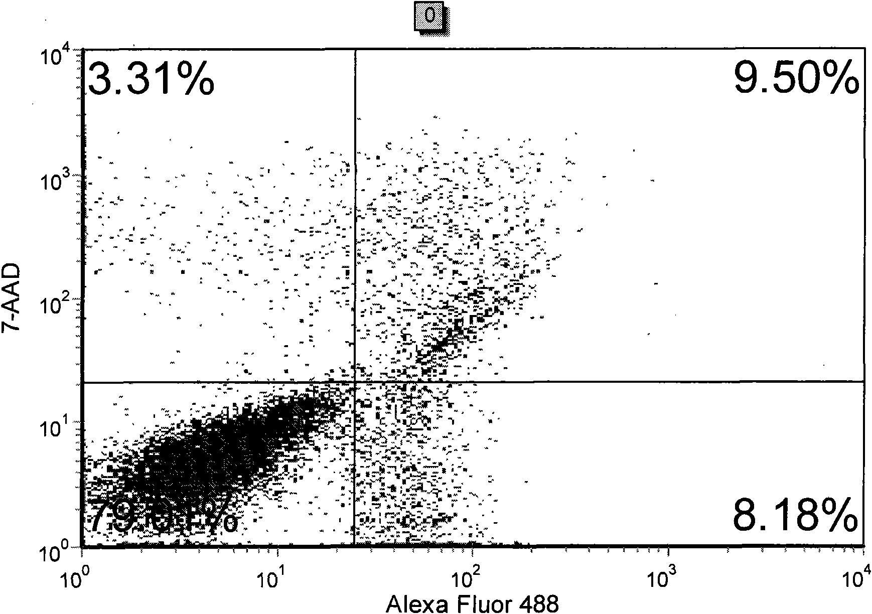
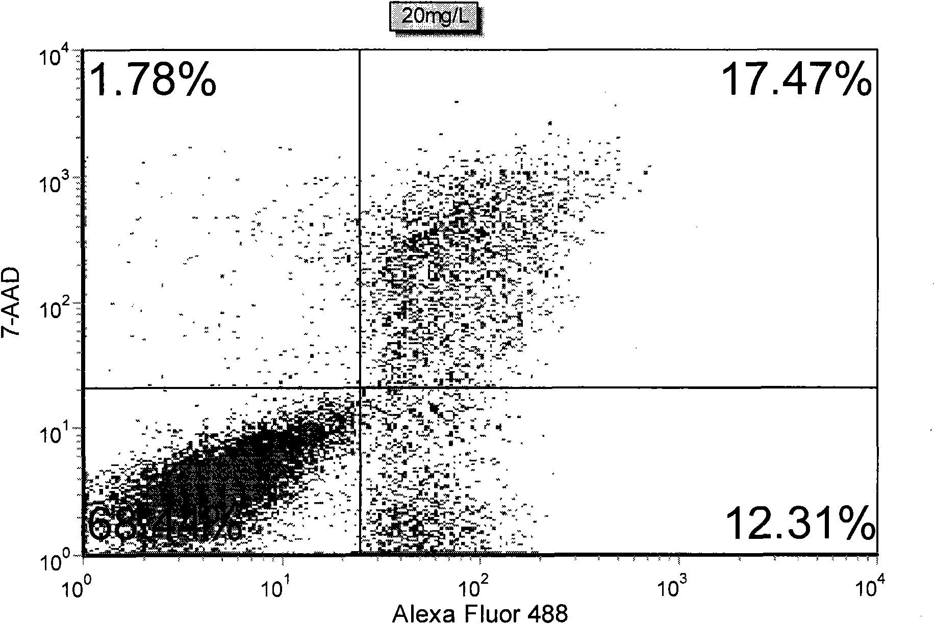
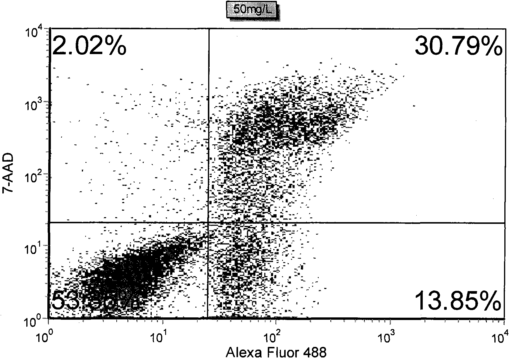
[0017] This example is an example of the detection of early apoptosis of human leukemia T lymphocyte line (Jurkat cell) by using the present invention.

[0018] Human leukemia T lymphocyte line (Jurkat suspension cells), grown in RPMI-1640 complete culture medium containing 10% fetal bovine serum, 100U / ml penicillin-streptomycin, placed at 37°C, 5% CO 2 cultured in an incubator. Adjust the density of the Jurkat suspension cell suspension in the logarithmic growth phase to 2.5×10 5 cells / ml, then pipette 3ml of cell suspension into a 60mm sterile petri dish, and add the apoptosis-inducing drug EGCG to each petri dish so that the final concentrations were 20, 50, 80, 100 and 120mg / ml respectively. L, at the same time set cells without drug treatment as a blank control, and then put them in a 37°C incubator for static culture for 24 hours.

[0019] Collect the Jurkat cells treated with EGCG for 24 hours into a centrifuge tube, centrifuge at 1000rpm for 10 minutes, remove the su...

Embodiment 2

[0023] This example is an example of the detection of early apoptosis of human liver cancer SMMC-7721 by using the present invention.

[0024] Human liver cancer SMMC-7721 cells were grown in RPMI-1640 complete culture medium containing 10% fetal bovine serum, 100U / ml penicillin-streptomycin, placed at 37°C, 5% CO 2 cultured in an incubator. Digest SMMC-7721 cells in the logarithmic growth phase into a cell suspension, and adjust the density to 2.5×10 5 cells / ml, and then use a pipette to draw 3ml of cell suspension into a 60mm sterile Petri dish. After culturing overnight and waiting for the cells to adhere to the wall, add the apoptosis-inducing drug EGCG to each culture dish to make the final concentrations of 50, 100, 125, 150 and 175 mg / L respectively, and set the cells without drug treatment as a blank control. Then put them in a 37°C incubator and culture them for 24 hours.

[0025] Collect the SMMC-7721 cells after 24 hours of EGCG into a centrifuge tube, centrifuge a...


PUM


Abstract

The invention discloses a method for detecting early apoptosis of cells. A phosphatidylserin antibody marked with a fluorescent dye Alexa Fluor 488 and a nucleic acid fluorescent dye 7-AAD are matched to detect the cells together, wherein the phosphatidylserin antibody is used for identifying normal living cells and apoptotic cells, and the nucleic acid fluorescent dye is used for identifying early apoptotic cells and late apoptotic cells in the apoptotic cells. After the two fluorescent dyes, namely the Alexa Fluor 488 and the 7-AAD with different emission wavelengths are detected and analyzed through a flow cytometry, the cells are divided into four subsets, wherein the Alexa Fluor 488+ / 7-AAD- subset is the early apoptotic cells.

Description

technical field [0001] The invention belongs to the field of biotechnology, and relates to a detection method for cell apoptosis, in particular to a detection method capable of distinguishing early apoptotic cells and late apoptotic cells. Background technique [0002] Apoptosis, or programmed cell death (PCD), refers to the active and physiological death process of the cell itself according to its own program under certain physiological or pathological conditions. It is a multi-step process. , the occurrence process of gene regulation involves the activation, expression and regulation of a series of genes. Apoptosis plays an important role in embryonic development and morphogenesis, the stability of normal cell populations in tissues, the defense and immune response of the body, cell damage and aging caused by disease or poisoning, and the occurrence and development of tumors, and has potential therapeutic significance . In the 1990s, the study of apoptosis was suddenly p...

Claims


Application Information

Patent Timeline
Patent Type & Authority Applications(China)
IPC IPC(8): G01N21/64C12Q1/68C12Q1/02
Inventor 何敏李刚钟艳平萧浩
Owner GUANGXI MEDICAL UNIVERSITY
Learn More