Electrochemical detection method for DNA three-dimensional nanostructure probe

A three-dimensional nano- and nano-structure technology, applied in biochemical equipment and methods, microbial determination/inspection, measuring devices, etc., can solve the problems of strict control of probe distance and unfavorable DNA biological activity, etc., to achieve high selectivity, Effects to avoid interactions and improve performance

Active Publication Date: 2010-12-22
SHANGHAI INST OF APPLIED PHYSICS - CHINESE ACAD OF SCI
View PDF2 Cites 23 Cited by
  • Summary
  • Abstract
  • Description
  • Claims
  • Application Information

AI Technical Summary

Problems solved by technology

Although this method controls the distance between the probes to a certain extent, and at the same time ensures that the probe DNA has a certain degree of freedom on the surface, in practice, this method controls the distance between the probe DNA by mixing self-assembly layers. The statistical method of the distance cannot strictly control the distance between the probes on the nanoscale, because molecules with similar groups tend to combine together, which is easy to form aggregation effects, which is not conducive to the biological activity of DNA

Method used


Image

  • Electrochemical detection method for DNA three-dimensional nanostructure probe
  • Electrochemical detection method for DNA three-dimensional nanostructure probe
  • Electrochemical detection method for DNA three-dimensional nanostructure probe

Examples


Embodiment 1

[0042] Reagents include:

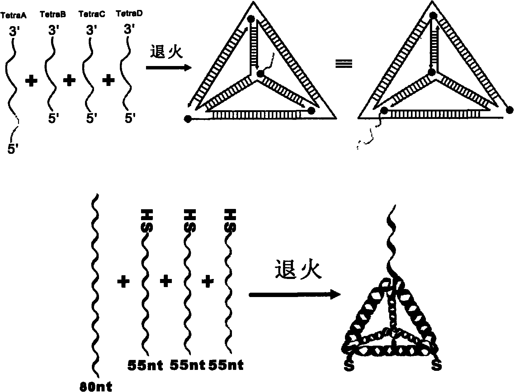
[0043] 4 pieces of single-stranded DNA used to assemble tetrahedral DNA nanostructure probes, Tetra-A (80bp, molecular weight 24539.0, ssDNA), Tetra-B (55bp, molecular weight 17018.0, 5' end modified sulfhydryl ssDNA), Tetra-C (55bp, molecular weight 16898.0, ssDNA modified with thiol at the 5' end), Tetra-D (55bp, molecular weight 16877.0, ssDNA modified with thiol at the 5' end), both were purchased from Dalian Takara Biological Co., Ltd.

[0044] The four DNAs that constitute the tetrahedral structure probe contain three structural domains, each of which is complementary to the corresponding domains of the other three single-stranded DNAs (17 pairs of bases are complementary), and each single-stranded DNA surrounds the tetrahedral structure One side of a circle, each vertex contains two bases (non-complementary, flexible) for bending, the 3' end and 5' end of the single-stranded DNA converge at the four vertices of the tetrahedron, and TetraA is a...

Embodiment 2

[0080] Reagents include:

[0081] DNA is the same as Example 1

[0082] Avidin-modified glucose oxidase (GOx-A), purchased from Vector Laboratories (SanDiego, CA)

[0083] The experimental procedure is the same as that in Example 1, except that the avidin-modified horseradish peroxidase is replaced by avidin-modified glucose oxidase, and the substrate TMB is replaced by glucose and benzoquinone. Voltage is 0.35V (reference is silver / silver chloride electrode), all the other parameters are constant, the result is the same as that of Example 1.

Embodiment 3

[0085] Reagents include:

[0086]4 pieces of single-stranded DNA used to assemble tetrahedral DNA nanostructure probes, S1 (88bp, molecular weight 27014.0, ssDNA), S2 (83bp, molecular weight 25642.6, ssDNA modified with thiol at the 5' end), S3 (83bp, molecular weight 25678.6, 5' end modified thiol ssDNA), S4 (83bp, molecular weight 25535.5, 5' end modified thiol ssDNA), were purchased from Dalian Takara Biological Co., Ltd. S1:

[0087] 5'-GTATCCAGTGGCTCATTTTTTTTTACGAACATTCCTAAGTCT

[0088] GAAATTTATCACCCGCCATAGTAGACGTATCACCAGGCAGTTG

[0089] AG-3'

[0090] S2:

[0091] 5’-HS-ATTCAGACTTAGGAATGTTCGACATGCGAGGAGGAAATG

[0092] AAGTCCAATACCGACGATTACAGGCCTTTGCGCCTTGCTACAC

[0093] G-3'

[0094] S3:

[0095] 5’-HS-ACGTGTAGCAAGGCGCAAAGGCCTGTAATCGACTCTAC

[0096] GGGAAGAGCATGCCCATCCGGCTCACTACTATGGCGGGTGAT

[0097] AAA-3'

[0098] S4:

[0099] 5’-HS-ACTCAACTGCCTGGTGATACGAGAGCCGGATGGGCATG

[0100] CTCTTCCCGTAGAGACGGTATTGGACTTCATTTTCCTCCTCGCA

[0101] TG-3'

[0102] in,

...


PUM

PropertyMeasurementUnit
diameteraaaaaaaaaa

Abstract

The invention discloses an electrochemical detection method for a DNA three-dimensional nanostructure probe, which sequentially comprises the following steps of: 1) synthesizing the DNA three-dimensional nanostructure probe by a self-assembly method, wherein the probe comprises an extended section of recognition sequence; 2) assembling the nanostructure probe onto the surface of a working electrode of an electrochemical device; 3) hybridizing target DNA and the probe on the surface of the working electrode; and 4) adding enzyme and a corresponding substrate for redox reaction and performing electrochemical detection by using the electrochemical device.

Description

technical field [0001] The invention belongs to the field of gene hybridization detection, and specifically relates to an electrochemical detection method using a DNA three-dimensional nanostructure probe. Background technique [0002] DNA electrochemical biosensors have huge potential applications in various fields such as clinical medicine, food inspection, environmental monitoring and anti-terrorism due to their advantages of fast, sensitive, low cost and easy miniaturization. An important aspect that determines the performance of DNA electrochemical biosensors is the molecular recognition interface. DNA biomolecules attached to the interface will inevitably reduce its biological activity due to its disordered orientation, reduction of free energy and strong interaction with the interface. In order to maximize the performance of biosensors, we need to find a way that does not hinder the active attachment of biomolecules to the surface as much as possible. [0003] The i...

Claims


Application Information

Patent Timeline
Patent Type & Authority Applications(China)
IPC IPC(8): C12Q1/68C12Q1/28C12Q1/26G01N27/327
Inventor 樊春海裴昊
Owner SHANGHAI INST OF APPLIED PHYSICS - CHINESE ACAD OF SCI
Why Eureka
Try Eureka