Method for preparing aqueous gel/nano hydroxyapatite composite scaffold for hard tissue repair

A technology of hydroxyapatite and composite scaffold, which is applied in the field of biomedical material science, can solve the problems of difficult tissue penetration and insufficient material performance, and achieve the effects of good induction of bone formation, improvement of bone repair effect, and obvious effect

Inactive Publication Date: 2011-01-26
TONGJI UNIV
View PDF5 Cites 17 Cited by
  • Summary
  • Abstract
  • Description
  • Claims
  • Application Information

AI Technical Summary

Problems solved by technology

The innovative design of material selection and preparation process solves the shortcomings of insufficient material performance, difficult formation of vascularization, and difficulty of tissue penetration into the stent.

Method used


Image

  • Method for preparing aqueous gel/nano hydroxyapatite composite scaffold for hard tissue repair
  • Method for preparing aqueous gel/nano hydroxyapatite composite scaffold for hard tissue repair
  • Method for preparing aqueous gel/nano hydroxyapatite composite scaffold for hard tissue repair

Examples


Embodiment 1

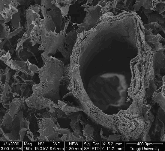
[0026] Step 1: Dissolve collagen and PLA with a concentration of 8% in hexafluoroisopropanol at a mass ratio of 1:2. After completely dissolving, perform electrospinning. The spinning parameters are: voltage 18kv, speed 500r / min , with a height of 10 cm, prepare small blood vessels with a diameter of 0.5 mm, cut into small sections with a length of 2 cm, and set aside;

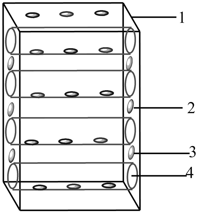
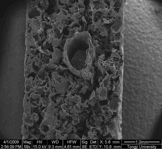
[0027] Step 2: Mix PLA and PCL at a mass ratio of 1:1, add sodium chloride after dissolving chloroform, let it stand for 1 hour, filter with salt to make holes, add small blood vessels and heat press to form a degradable three-dimensional scaffold with reserved channels Material;

[0028] Step 3: Dissolve 1g of degradable water-soluble polymer PEG2000 in 10ml of water, dissolve 0.1g of α-cyclodextrin and 0.005g of nano-hydroxyapatite in 10ml of water, ultrasonically disperse for 15 minutes, and mix the two solutions by 1: 1 volume ratio mixed, immediately injected into the reserved channel of the scaffold mat...

Embodiment 2

[0032] Step 1: Dissolve collagen and PLA with a concentration of 8% in hexafluoroisopropanol at a mass ratio of 1:3. After completely dissolving, electrospinning is performed. The spinning parameters are: voltage 16kv, speed 600r / min , with a height of 12 cm, prepare small blood vessels with a diameter of 1.0 mm, cut into small sections with a length of 2 cm, and set aside;

[0033] Step 2: Mix PLA and PCL at a mass ratio of 1:1, add sodium chloride after dissolving chloroform, let it stand for 1 hour, filter with salt to make holes, add small blood vessels and heat press to form a degradable three-dimensional scaffold with reserved channels Material;

[0034] Step 3: Dissolve 1g of degradable water-soluble polymer PEG2000 in 10ml of water, dissolve 0.1g of α-cyclodextrin and 0.005g of nano-hydroxyapatite in 10ml of water, ultrasonically disperse for 15 minutes, and mix the two solutions by 1: 1 volume ratio mixed, immediately injected into the reserved channel of the scaffol...

Embodiment 3

[0038] Step 1: Dissolve collagen and PLA with a concentration of 8% in hexafluoroisopropanol at a mass ratio of 1:2. After completely dissolving, perform electrospinning. The spinning parameters are: voltage 18kv, speed 500r / min , with a height of 10 cm, prepare small blood vessels with a diameter of 0.5 mm, cut into small sections with a length of 2 cm, and set aside;

[0039] Step 2: Mix PLA and PCL at a mass ratio of 1:2, add sodium chloride after dissolving chloroform, let it stand for 1 hour, filter with salt to make holes, add small blood vessels and heat press to form a degradable three-dimensional scaffold with reserved channels Material;

[0040] Step 3: Dissolve 1g of degradable water-soluble polymer PEG-PCL-PEG in 10ml of water, dissolve 0.1g of α-cyclodextrin and 0.005g of nano-hydroxyapatite in 10ml of water, and ultrasonically disperse for 15 minutes. The solution is mixed according to a volume ratio of 1:1, and immediately injected into the reserved channel of th...


PUM


Abstract

The invention belongs to the field of bio-medicinal material science, and particularly relates to a method for preparing an aqueous gel/nano hydroxyapatite composite scaffold for hard tissue repair. The method comprises the following steps of: preparing a small blood vessel by an electrostatic spinning method; preparing a degradable three-dimensional scaffold material taking the small blood vessel as a reserved passage inner wall by using a hot pressing/salting-out method; injecting injectable super-molecular aqueous gel prepared from cyclodextrin, tri-block polymer and nano hydroxyapatite into the reserved passage; and soaking the obtained scaffold into growth factor culture liquid for modification. The composition, property and shape of the prepared scaffold material are more suitable for bone surface blood vessel formation, and new cells and nutrients can enter the inner side of the scaffold through the aqueous gel in the reserved passage to induce bone growth so as to better finish the repair of bone defects. The bone repair effect of the scaffold material is obviously improved compared with other bone repair materials, and the mechanical property, blood vessel function and bone repair function of the scaffold material are obvious compared with common bone repair materials.

Description

technical field [0001] The invention belongs to the field of biomedical material science, and in particular relates to a method for preparing a hydrogel / hydroxyapatite composite bracket for hard tissue repair. Background technique [0002] In 1993, Langer and Vacanti proposed that the basic meaning of tissue engineering is to apply the basic principles and technologies of engineering and life sciences to construct biologically functional substitutes in vitro for repairing tissue defects, replacing functional or exhausted tissues, Part or all of the function of an organ. Bone tissue engineering scaffold material is a discipline that researches and develops biological substitutes for repairing and improving the shape and function of damaged bone tissue. On the one hand, bone tissue engineering serves as the carrier of seed cells and growth factors to transport them to the defect site, and on the other hand, it also provides the function of forming new bone. A good bone scaffo...

Claims


Application Information

Patent Timeline
Patent Type & Authority Applications(China)
IPC IPC(8): A61L27/14A61L27/20A61L27/12A61L27/54A61L27/24A61L27/18A61F2/28
Inventor 李兰冯玥任天斌
Owner TONGJI UNIV
Why Eureka
Try Eureka