Breast mass and calcific benign-malignant automatic recognition and quantitative image evaluation system

A technology of automatic identification and evaluation system, applied in the field of medical diagnostic equipment for breast diseases, can solve the problem of lack of quantitative evaluation indicators, achieve the effect of convenient operation and improve the accuracy of judgment

Inactive Publication Date: 2011-02-16
SUN YAT SEN UNIV
View PDF3 Cites 14 Cited by
  • Summary
  • Abstract
  • Description
  • Claims
  • Application Information

AI Technical Summary

Problems solved by technology

[0005] The purpose of the present invention is to overcome the problem of the lack of quantitative evaluation index in the existing mammography film (nuclear magnetic field, ultrasound) image diagnosis, and to provide a quantitative image-aided diagnosis evaluation system for mammary gland lesions. Quantitative analysis of target film (MRI, ultrasound) images to provide predictive value for benign and malignant breast lesions

Method used


Image

  • Breast mass and calcific benign-malignant automatic recognition and quantitative image evaluation system
  • Breast mass and calcific benign-malignant automatic recognition and quantitative image evaluation system
  • Breast mass and calcific benign-malignant automatic recognition and quantitative image evaluation system

Examples


Embodiment 1

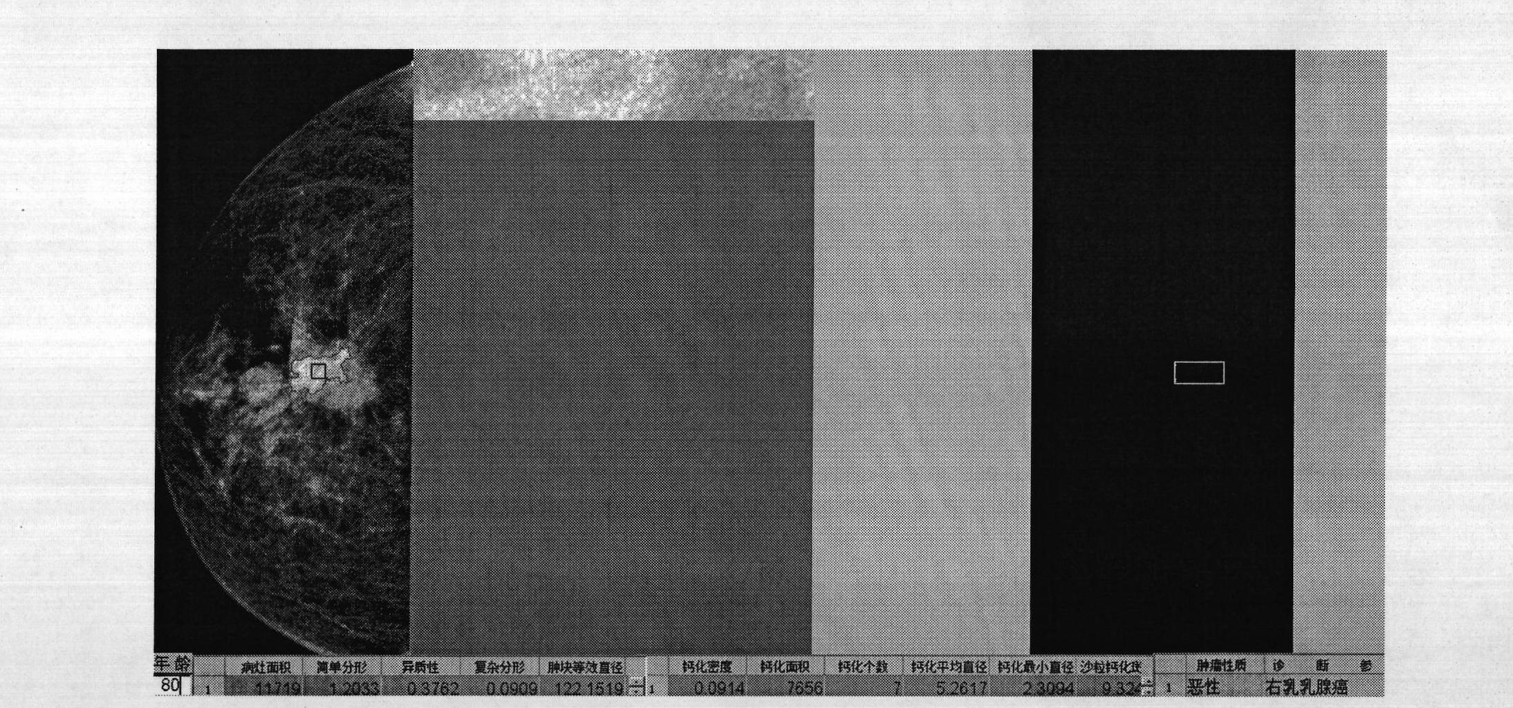
[0037] Patient A; age: 80 years old; clinical diagnosis: grade 2 malignant breast cancer without nipple discharge

[0038] Use the CAD system to automatically obtain the ROIs of benign and malignant tumors in mammography. Figure 1A , Figure 1B with Figure 1C They are examples of tumors automatically identified by mammography. The left picture in the figure is the original mammogram. The area surrounded by the red curve is the malignant mass automatically recognized by CAD. The middle picture combined with the left picture is the enlarged calcified area. Screenshot, the right picture is the calcified plaque automatically extracted by CAD, and the yellow rectangle is the circumscribed rectangle of the calcified area. When no yellow rectangle box appears automatically, it indicates that there is no calcified plaque in the image (such as Figure 1C ), once the yellow rectangular frame appears automatically, it indicates the existence of microcalcified plaques, even if no ob...

Embodiment 2

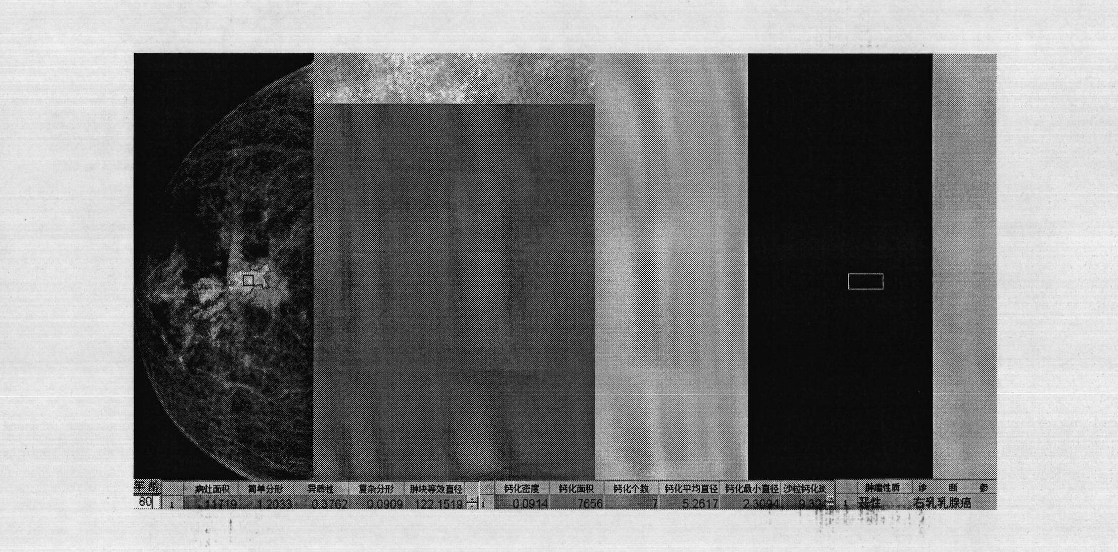
[0042] Patient B; age: 75 years old; clinical diagnosis: grade 3 malignant breast cancer with nipple discharge

[0043] Use the CAD system to automatically obtain the ROIs of benign and malignant tumors in mammography. Processed graphics are similar to those described in Example 1 Figure 1A . The left picture is the original mammography target film, the middle is the enlarged picture of the malignant calcified plaque automatically identified by CAD, and the yellow rectangle is the smallest circumscribed rectangle of the calcified plaque. All the image parameter values ​​mined and extracted are located at the bottom area of ​​the CAD system graphical interface, and the evaluation reference obtained based on the built-in model is located at the bottom right corner of the CAD system graphical interface.

[0044] Calculated from CAD Figure 1B Mass parameter in: fractal dimension D F = 1.2459, heterogeneity H = 0.4735, multifractal dimension M F =0.1364, calcification param...

Embodiment 3

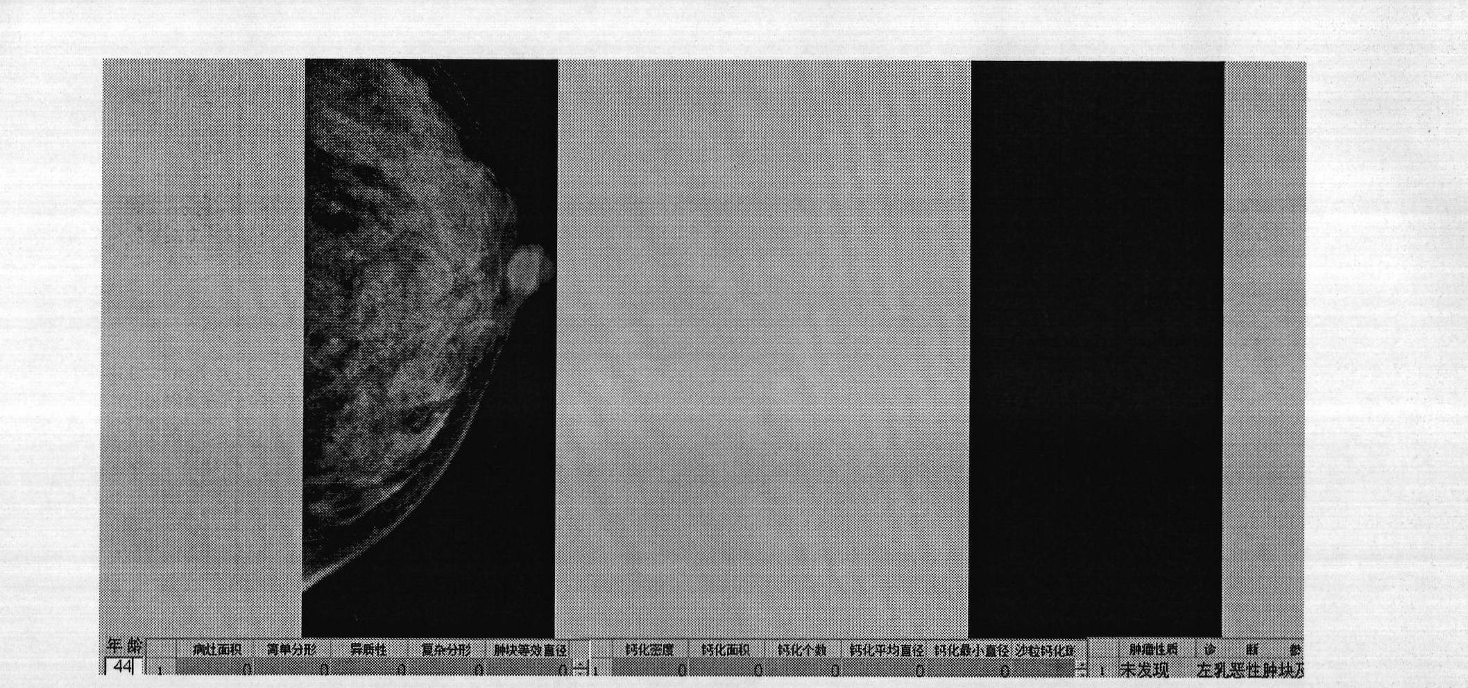
[0047] Patient C; age: 44 years old; clinical diagnosis: mastitis with tenderness

[0048] Calculated from CAD Figure 1C Mass parameter in: fractal dimension D F = 0, heterogeneity H = 0, multifractal dimension M F =0, calcification parameters: calcification density P=0, number of calcification spots N=0, sand calcification spots Ns=0,

[0049] Since the results of the CAD examination showed no lumps and calcified plaques, the calculation results were all 0, and the judgment result was that no malignant lumps and calcifications were found, which was consistent with the clinical diagnosis (mastitis is only a common general breast disease)

[0050] Judgment regression equation (1) according to classification: Y E =a*D F +b*M F +c*y+d*P+e*H, according to the logic equation (5) for distinguishing good and bad tumors: E=M mul |F simp |y*P&Ns, joint analysis discriminant. Based on the automatic analysis and judgment of the above CAD system, Figure 1C It was judged by CA...


PUM


Abstract

The invention provides a breast lesion quantitative image evaluation (CAD) system and an application method thereof. The breast lesion quantitative image evaluation system adopts image data of a fractal technology, pattern analysis, and the like to extract an excavating mean and a mathematical modeling algorithm, combines clinical data to establish a breast lesion grown diffusion nonlinear data model and is applied to tumor medical image analysis and tumor disease risk evaluation. The nonlinear data model comprises the clinical parameters of breast tumor grown diffusion state characteristic parameters, calcific state characteristic parameters, breast surface characteristic limitation asymmetric compaction comparison, nipple retraction, pachyderma, structural distortion, and the like. The breast lesion quantitative image evaluation system has a full-graphical interface, can lead a molybdenum target, nuclear magnetism and ultrasonic image data in and is convenient and quick in operation(one-key operation). By the CAD system, a benign-malignant forecasting numerical value and a tumor classification forecasting value of breast molybdenum target piece (nuclear magnetism and ultrasonic) lesions can be calculated, and results of the benign-malignant forecasting numerical value and the tumor classification forecasting value can be applied to breast image auxiliary diagnosis and breast filming screening.

Description

technical field [0001] The invention belongs to a medical diagnostic device for breast disease, and in particular relates to an automatic identification and quantitative image (mammography, ultrasound, NMR) evaluation system for breast lumps and calcifications, including image data segmentation and extraction and image data based on clinical pathology data. mathematical modeling; the system of the present invention is suitable for general clinical auxiliary diagnosis and general survey. Background technique [0002] Breast imaging examinations, especially mammography, provide an important basis for the diagnosis, staging, and efficacy evaluation of breast tumors, and have been used in breast screening. Unfortunately, in the current clinical diagnosis, routine imaging diagnosis is limited to the measurement of tumor size and simple shape factor and some qualitative evaluations, even some doctors or medical practitioners who can use the foreign image processing software Image-...

Claims


Application Information

Patent Timeline
Patent Type & Authority Applications(China)
IPC IPC(8): G06F19/00
Inventor 邵元智李立范家远
Owner SUN YAT SEN UNIV
Why Eureka
Try Eureka