Nano structure for enhancing Raman and fluorescence signals and preparation method thereof

A technology of nanostructure and fluorescent signal, which is applied in the field of nanostructure and its preparation to enhance Raman and fluorescent signal, can solve the problems that cannot meet the needs of rapid and ultra-sensitive detection of nucleic acid, shorten the detection time, improve the detection signal, and ensure The effect of gap stabilization

Inactive Publication Date: 2011-05-18
SOUTHEAST UNIV
View PDF2 Cites 4 Cited by
  • Summary
  • Abstract
  • Description
  • Claims
  • Application Information

AI Technical Summary

Problems solved by technology

The research by Nam et al. has realized the interstitial positioning of signal molecules (dye), but after hybridization with target molecules, silver deposition is performed, and the Raman signal can only be measured by exploring the thickness of the silver shell, which cannot meet the needs of rapid and ultra-sensitive detection of nucleic acids in actual samples.

Method used


Image

  • Nano structure for enhancing Raman and fluorescence signals and preparation method thereof
  • Nano structure for enhancing Raman and fluorescence signals and preparation method thereof
  • Nano structure for enhancing Raman and fluorescence signals and preparation method thereof

Examples


Embodiment 1

[0022] Example 1: Preparation method of gold nanostructures that enhance Raman and fluorescence signals

[0023] Adjust the gold nanoparticle suspension with a particle size of 50 nm to a concentration of 1 × 10 6-18 ions per liter, the ions adsorbed on the surface of metal nanoparticles will form a stable electric double layer structure, and the particles repel each other due to the electrostatic interaction of the stable electric double layer structure. Then slowly drop the diluted functionalized particle size 8 nm ferrite magnetic nanoparticles in the ratio of 1:2-50 (number) in the gold nanoparticle suspension (the method used for functionalization is known in the art of). A small amount of magnetic nanoparticles is in an excess of gold nanoparticle suspension, thereby ensuring multiple gold nanoparticles around one magnetic nanoparticle rather than multiple magnetic nanoparticles around one gold nanoparticle. Magnetic nanoparticles are adsorbed to the surface of gold na...

Embodiment 2

[0024] Example 2: Preparation method of gold-shell nanostructures that enhance Raman and fluorescence signals

[0025] Adjust the nanoparticle suspension of the silica core gold shell structure with a particle size of 180 nm to a concentration of 1×10 10 per liter, the ions adsorbed on the surface of gold-shell nanoparticles will form a stable electric double layer structure, and the particles repel each other due to the electrostatic interaction of the stable electric double layer structure. Slowly add diluted functionalized ferrite magnetic nanoparticles with a particle size of 10 nm to the gold nanoparticle suspension at a ratio of 1:10 (number) (the method used for functionalization is known in the art) . A small amount of magnetic nanoparticles is in an excess of gold nanoparticle suspension, thereby ensuring that there are multiple gold-shell nanoparticles around one magnetic nanoparticle rather than multiple magnetic nanoparticles around one gold-shell nanoparticle. M...

Embodiment 3

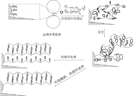
[0026] Embodiment 3: the application of the gold nanostructure that enhances Raman and fluorescent signal in detection——nucleic acid detection (Raman signal)

[0027] Add 10 μl of carboxyl-activated nucleic acid probe molecules (10 nmol / L) to 50 μl of gold nanostructures (1×10 8 per liter), overnight at 4°C to wash away unreacted nucleic acid probe molecules. Add the solution to be tested to the probe-immobilized gold nanoshell nanostructure suspension, hybridize at 72°C for 5 minutes, and then heat cycle for 3 times (60°C / 4°C). Then add single-stranded nuclease hydrolase to treat for 5 minutes at 37°C, wash and separate the gold nanostructures with a magnetic field, and measure the characteristic signal of nucleic acid with a Raman spectrometer (the experimental process is shown in Figure 3). The methods used are known in the art and can be utilized.


PUM

PropertyMeasurementUnit
Particle sizeaaaaaaaaaa

Abstract

The invention discloses a uniform nano aggregate and a preparation method. The nano aggregate comprises a magnetic nano particle modified with sulfydryl, wherein two sides of the magnetic nano particle are respectively connected with a first metal nano particle and a second metal nano particle through the sulfydryl, the first metal nano particle, the magnetic nano particle and the second metal nano particle are arranged in a line, the particle sizes of the first and the second metal nano particles are 20-400nm and the first and the second metal nano particles are same metal nano particles, and the particle size of the magnetic nano particle is 3-20nm. An object to be tested is orientated to a gap between the metal nano particles by using function groups on the surfaces of the nano particles, thus the great enhancement of the Raman and fluorescence signals of a molecular to be tested is realized. The invention can provides a novel rapid and ultra-sensitive detection method for experiment research, clinical diagnosis and large sample screening.

Description

technical field [0001] The invention relates to the technical field of functional nanomaterials, more specifically, a nanostructure for enhancing Raman and fluorescence signals and a preparation method thereof. Background technique [0002] In today's life science research, the understanding and resolution of major issues such as the law of life activities and the occurrence and development of diseases have entered the study of single-molecule level. The development and application of new molecular-level detection methods has become a research hotspot in the field of medical biology today. It not only promotes revolutionary changes in health-related diagnostic methods, but also has practical implications in biological and medical research, drug development, and disease prevention and treatment. significance. [0003] Existing highly sensitive nucleic acid detection methods for specific sequences are generally based on polymerase chain reaction (PCR) and fluorescent labeling...

Claims


Application Information

Patent Timeline
IPC IPC(8): C09K11/58C09K11/59C09K11/02B82Y25/00B82Y40/00
Inventor 董健许蓓蓓陶琴钱卫平
Owner SOUTHEAST UNIV
Why Eureka
Try Eureka