Preparation method of trachea substitute

A substitute and trachea technology, applied in the field of preparation of natural decellularized trachea substitutes, can solve problems such as failure to effectively remove cartilage ring antigen components, inability to achieve tracheal reconstruction, failure of tracheal reconstruction, etc., achieving good integration effect and source of raw materials Low cost, lower medical costs

Active Publication Date: 2011-05-25
西安博鸿生物技术有限公司
View PDF7 Cites 4 Cited by
  • Summary
  • Abstract
  • Description
  • Claims
  • Application Information

AI Technical Summary

Problems solved by technology

The author of the article also admits that the chondrocytes in the porcine tracheal tissue cannot be removed according to the method he introduced, and it can be observed that intact chondrocytes exist in the cartilage ring structure of the decellularized tracheal scaffold; subsequently, in the process of repairing the dog tracheal defect , the cartilage ring structure of the trachea has obvious absorption. With the extension of implantation time, the cartilage ring will be further absorbed and degraded, and the effect of tracheal reconstruction cannot be achieved.
It can be seen that the decellularization method of porcine tracheal tissue in the article failed to effectively remove the antigenic components in the cartilage ring, which may cause immune rejection of the recipient, resulting in degradation of the cartilage ring and failure of tracheal reconstruction

Method used


Image

  • Preparation method of trachea substitute
  • Preparation method of trachea substitute
  • Preparation method of trachea substitute

Examples


Embodiment 1

[0025] Step 1. De-antigen treatment of animal trachea

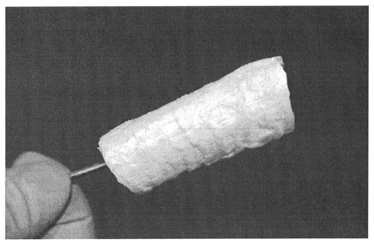
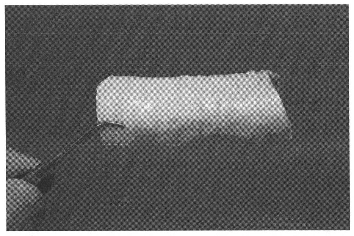
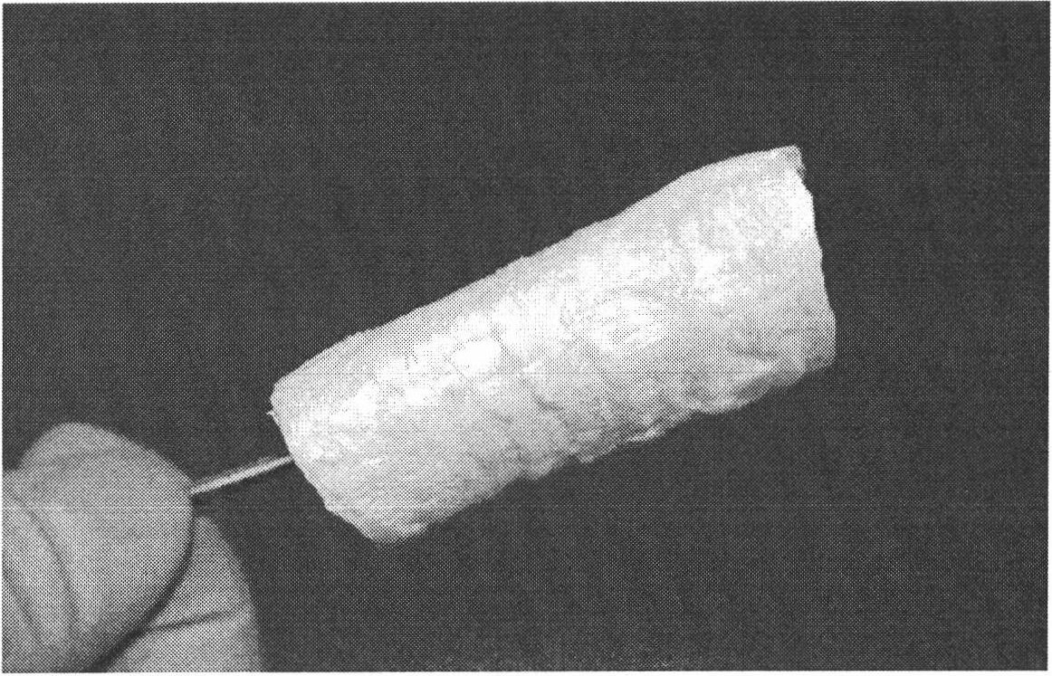
[0026] Raw material pretreatment: intercept 10 cm of goat tracheal tissue, remove surface blood stains and accompanying soft tissues, and obtain tracheal tissue with cartilage ring structure; wash with PBS solution for 3 times; (the connective tissue surrounded by the outer wall of tracheal tissue must be removed thoroughly, which will affect Transplanted trachea and surrounding tissue to establish blood supply).

[0027] Degreasing treatment: put the pretreated tracheal tissue into ether solution, ultrasonically oscillate for 30 minutes, and rinse with running water for 3 hours; (ether has a degreasing effect, and ultrasonic treatment can speed up the dissolution of free lipid droplets).

[0028] Antigen removal treatment on the inner wall: seal both ends of the degreased tracheal tissue, add trypsin solution with a m / v concentration of 0.2% in the tracheal lumen before sealing, and replace it with NaCl with a m / v concentr...

Embodiment 2

[0036] Step 1. De-antigen treatment of animal trachea

[0037] Raw material pretreatment: cut out 7 cm of porcine tracheal tissue, remove surface blood stains and accompanying soft tissues, obtain animal tracheal tissue with cartilage ring structure, wash with PBS solution for 3 times;

[0038] Degreasing treatment: Put the pretreated tracheal tissue into ether solution, treat it with ultrasonic vibration for 1 hour, and wash it with running water for 3 hours;

[0039] Inner wall anti-antigen treatment: seal both ends of the degreased tracheal tissue, add trypsin solution with a m / v concentration of 0.1% in the tracheal lumen before sealing, and replace it with NaCl with a m / v concentration of 2% after shaking for half an hour The solution was shaken and washed for half an hour, and then replaced with a NaCl solution with a m / v concentration of 0.9% for half an hour;

[0040] Antigen removal treatment on the outer wall: place the tracheal tissue that has been treated with ant...


PUM


Abstract

The invention provides a preparation method of a trachea substitute. The preparation method comprises the following steps: eliminating antigens of an animal-derived tracheal tissue; compounding vascular endothelial growth factors (VEGF) with the outer wall of the trachea substitute; and compounding epidermal growth factors (EGF) with the inner wall of the substitute, wherein antigen elimination is carried out on a cartilage layer on the outer wall of a trachea and a lower muscular layer of a mucous layer on the inner wall of the trachea. The trachea substitute prepared by the method is derived from the animal tracheal tissue, has a specific C-shaped cartilaginous ring structure of a natural trachea, is formed by connecting compact connective tissues, has elasticity, can extend and contract longitudinally, and can moderately expand when gas passes through the substitute; and the prepared trachea substitute can keep the trachea unobstructed for a long time after transplantation, can notcollapse, and has good integration effect with a host without obvious rejection reaction. The preparation method has the advantages of simpleness, wide raw material source and low cost; and the medical cost can be reduced and the economic burden of patients can be lightened by means of large-scale industrial production.

Description

technical field [0001] The invention belongs to the technical field of tissue engineering medical biomaterials, and in particular relates to a preparation method of a natural decellularized trachea substitute. Background technique [0002] Tracheal stenosis caused by diseases such as tracheal tumors and tracheal scars can cause severe breathing difficulties and even death by suffocation. Such tracheal lesions sometimes require resection of a segment of the trachea followed by tracheal reconstruction. When the length of the resected trachea exceeds 6 cm, a tracheal substitute or an allogeneic trachea is required to replace the original diseased trachea. Commonly used tracheal substitutes mainly include tracheal substitutes derived from autologous tissue, allogeneic trachea and artificial trachea. [0003] The sources of autologous tissue and allogeneic trachea are limited, and allogeneic trachea has the risk of immune rejection, while artificial trachea is difficult to have...

Claims


Application Information

Patent Timeline
Patent Type & Authority Applications(China)
IPC IPC(8): A61F2/04
Inventor 杨改叶
Owner 西安博鸿生物技术有限公司
Why Eureka
Try Eureka