Chitosan-DNA nanometer granule complex and preparation method thereof
A nanoparticle and chitosan technology, which can be used in pharmaceutical formulations, genetic material components, gene therapy, etc., can solve problems such as low encapsulation efficiency, poor storage stability, and application limitations, and achieve endocytosis promotion and low cytotoxicity Effect
- Summary
- Abstract
- Description
- Claims
- Application Information
AI Technical Summary
Problems solved by technology
Method used
Image

Examples
Embodiment 1
[0025] Embodiment one: the preparation of chitosan nanoparticle DNA complex CNPs-DNA
[0026] (1) Dissolve 20mg of purified chitosan in 10% acetic acid and heat it slightly (do not exceed 42°C). After the chitosan particles are completely dissolved, roughly adjust the pH to 5.5 with 10M NaOH solution to obtain 0.2 % (w / v) chitosan stock solution, sterilized by filtration through a 0.22 μm filter membrane.
[0027] (2) Take an appropriate amount of chitosan stock solution and dilute it into a chitosan solution with a concentration of 0.08% (w / v), adjust the pH to 6.5-7.0 with 10M NaOH solution, and filter through a 0.22μm filter membrane for future use .
[0028] (3) Dilute the plasmid DNA (pEGFP-C1) with distilled water to a DNA solution of 400 μg / ml, add 1 / 10 volume of 500mM Na 2 SO 4 solution (to a final concentration of 50mM).
[0029] (4) Chitosan solution and plasmid solution were placed in a constant temperature incubator at 55°C for 30 min. After mixing equal volum...
Embodiment 2
[0030] Embodiment 2: Determination of the physical properties of CNPs-DNA
[0031] 1. Gel electrophoresis retardation and DNase I digestion experiment: please specify the specific steps of gel electrophoresis retardation digestion experiment,
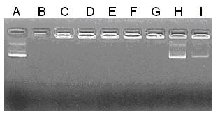
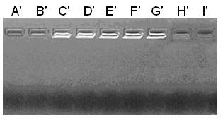
[0032] (1) Gel electrophoresis retardation experiment: CNPs-DNA was subjected to 1.0% agarose gel electrophoresis at 60V for 40min. Since the negative charge of the DNA itself was covered by the chitosan-wrapped DNA, it could not move from the negative pole to the negative pole in the electrophoresis. The positive electrode moves and is blocked in the loading hole.
[0033] (2) DNaseⅠdigestion experiment: add 0.5U DNaseⅠ (1U / ul) to CNPs-DNA, incubate at 37°C for 1h, electrophoresis on 1.0% agarose gel at 60V, 40min, the wrapped DNA stays in the sample well , the unencapsulated DNA was digested and degraded by DNase I. In this way, the protective ability of chitosan nanoparticles to DNA was judged. see figure 1 and figure 2 , indi...
Embodiment 3
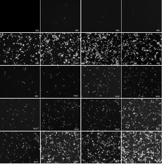
[0037] Example 3: Effect of CNPs-DNA with different N / P on transfection efficiency:
[0038] Apply CNPs-DNA to mediate the transfection of pEGFP-C1 to HEK293T cells, the specific steps are:
[0039] After recovery, the cells were subcultured, and the logarithmic growth phase was reached in about three generations (at this time, the cells grew vigorously and divided at the fastest speed), and the cells with good growth status were selected for testing. Count the cells after digesting them according to the previous cell passage steps, transfer them to a 24-well plate for culture, and the number of cells per well is about 5'105. After culturing for 16-24 hours, observe that the cells are uniformly attached to the wall and the density reaches 80%-90%, and then the transfection experiment can be performed.
[0040] (1) Remove the medium, carefully add PBS to wash the cells twice (do not damage or blow up the cells);
[0041] (2) Set up a blank control well, and only add complete ...
PUM
 Abstract
Description
Claims
Application Information
 - R&D
- Intellectual Property
- Life Sciences
- Materials
- Tech Scout
- Unparalleled Data Quality
- Higher Quality Content
- 60% Fewer Hallucinations
Browse by: Latest US Patents, China's latest patents, Technical Efficacy Thesaurus, Application Domain, Technology Topic, Popular Technical Reports.
© 2025 PatSnap. All rights reserved.Legal|Privacy policy|Modern Slavery Act Transparency Statement|Sitemap|About US| Contact US: help@patsnap.com