Duck viral enteritis virus infectious recombinant cloning system and its construction method and application

A technology for duck viral enteritis, infectivity, applied in the field of infectious cloning of virus vaccine strains

Active Publication Date: 2011-12-14
HARBIN VETERINARY RES INST CHINESE ACADEMY OF AGRI SCI
View PDF1 Cites 13 Cited by
  • Summary
  • Abstract
  • Description
  • Claims
  • Application Information

AI Technical Summary

Problems solved by technology

However, the research on the non-essential region for the replication of duck viral enteritis virus is still blank

Method used


Image

  • Duck viral enteritis virus infectious recombinant cloning system and its construction method and application
  • Duck viral enteritis virus infectious recombinant cloning system and its construction method and application
  • Duck viral enteritis virus infectious recombinant cloning system and its construction method and application

Examples


Embodiment 1

[0037] Example 1. Construction of Genome Fosmid Library of DEV Vaccine Strain

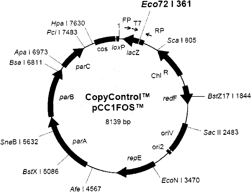
[0038] The Fosmid library of the DEV genome was constructed according to the instructions of the "CopyControl Fosmid Library Production Kit" kit from EPICENTRE. The method is as follows, the DNA of the DEV vaccine strain virus (CVCC AV1222) (GeneBank EU082088) (China Veterinary Microbiological Culture Collection Management Center, catalog number AV1222; purchased from the China Veterinary Drug Supervision Institute) DNA was physically used with a 25-gauge needle (purchased from Shanghai Zhiyu Medical Instrument Co., Ltd.) was pumped several times to cut the DNA fragments with T4DNA Polymerase (purchased from New England Biolabs) and alkaline phosphatase (Alkaline Phosphatase) (purchased from New England Biolabs). End smoothing and dephosphorylation treatment, pulse electrophoresis (with Bio-Rad CHEF The XA Pulsed Field system performs pulse electrophoresis, and the conditions of pulse electrophor...

Embodiment 2

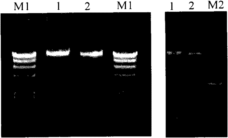
[0039] Example 2. Selection for rescue of DEV cosmids

[0040] After the library was successfully constructed, 286 clones were picked to extract the cosmids, and the alkaline lysis method was used to [5] Extract the cosmid and send it to Dalian Bao Biological Co., Ltd. to sequence the end of the DEV DNA fragment inserted into pCC1 Fos. The sequences of the sequencing primers are as follows:

[0041] Primer 1: TAATACGACTCACTATAGGG

[0042] Primer 2: GCCAAGCTATTTAGGTGAGA

[0043] After terminal sequencing analysis, a total of 250 clones with complete FseI-SbfI-PmeI adapters connected to both ends of the insert fragment were obtained. Multiple sets of 5 cosmid combinations were selected from these 250 clones. Both ends of the DEV DNA fragments cloned in each group contain Fse I-Sbf I-Pme I joints, which can overlap each other and can be spliced ​​to cover the entire DEV genome.

Embodiment 3

[0044] Example 3. Virus rescue

[0045] The DNA of the selected cosmids was extracted with Qiagen's medium extraction kit. The selected cosmids were linearized with Fse I, Sbf I or Pme I endonuclease (both purchased from New England Biolabs), and the reaction conditions were as follows: 20 U of Sbf I endonuclease (Fse I or Pme I could also be used Endonuclease), cosmid 10 μg, acted at 37°C for 1 hour, extracted with phenol / chloroform, and precipitated with ethanol to prepare DEV DNA for transfection.

[0046] Refer to the calcium phosphate method of Reddy SM (2002) to co-transfect the second generation chicken embryo fibroblasts (CEF) with 5 segments of DEV DNA [3] After multiple repetitions, 3 groups of 5-cosmid combinations showed typical lesions of DEV virus after 4-6 days of transfection. A group of 5-cosmid combinations with good repeatability was selected for subsequent experiments. The position of the cosmid combination relative to the DEV genome is as follows image...


PUM


Abstract

The invention relates to a duck enteritis virus infectious recombinant cloning system. The duck enteritis virus infectious recombinant cloning system is characterized by comprising a plurality of cosmids, wherein a duck enteritis virus vaccine strain gene segment is cloned in each cosmid, contains areas which are overlapped with each other, and a duck enteritis virus vaccine strain complete genome is assembled and covered; and an exogenous gene is inserted in a replication nonessential region of at least one cosmid. The invention also relates to a method for constructing the duck enteritis virus infectious recombinant cloning system and application of the duck enteritis virus infectious recombinant cloning system to the construction and recombination of the duck enteritis virus.

Description

technical field [0001] The present invention relates to the field of infectious cloning of virus vaccine strains, in particular to the field of infectious cloning of duck viral enteritis virus vaccine strains, more specifically to the infectious recombinant cloning system of duck viral enteritis virus vaccine strains, and its construction method and application . Background technique [0002] Duck enteritis virus (DEV), also known as duck plague virus. It can cause acute, febrile and septic acute infectious diseases in ducks, geese and other birds of Anseriformes. However, there are relatively few studies on DEV compared with other herpes viruses. Therefore, the eighth report of the International Commission on Classification of Virology classified it as a herpes virus [1] , without being able to classify it further. Until recently, its full sequence was not fully tested [2] (GeneBank EU082088). [0003] Since the use of poxviruses as live vaccine vectors in the 1980s, ...

Claims


Application Information

Patent Timeline
Patent Type & Authority Applications(China)
IPC IPC(8): C12N15/63C12N15/38C12N7/01A61K39/245A61P31/20C12R1/93
Inventor 陈化兰柳金雄步志高姜永萍
Owner HARBIN VETERINARY RES INST CHINESE ACADEMY OF AGRI SCI
Why Eureka
Try Eureka