Method for segmenting three-dimensional ultrasonic image

A technology of 3D ultrasound and ultrasound images, which is applied in image analysis, image enhancement, image data processing, etc. It can solve the problems of not using 3D spatial information, the difficulty of accurate segmentation of 3D ultrasound images, and the blurring of tissue boundaries and details.

Inactive Publication Date: 2012-04-04
SOUTH CHINA UNIV OF TECH
View PDF3 Cites 23 Cited by
  • Summary
  • Abstract
  • Description
  • Claims
  • Application Information

AI Technical Summary

Problems solved by technology

Among them, the processing methods based on two-dimensional space have the following disadvantages: (1) they do not use three-dimensional space information; (2) the obtained two-dimensional contour needs to be topologically connected and interpolated in three-dimensional space to reconstruct a high-precision and complete 3D image; (3) When calculating the volume, the interpolat

Method used


Image

  • Method for segmenting three-dimensional ultrasonic image
  • Method for segmenting three-dimensional ultrasonic image
  • Method for segmenting three-dimensional ultrasonic image

Examples


Embodiment Construction

[0064] The technical solutions of the present invention will be further described below in conjunction with the embodiments and the accompanying drawings, but the embodiments of the present invention are not limited thereto.

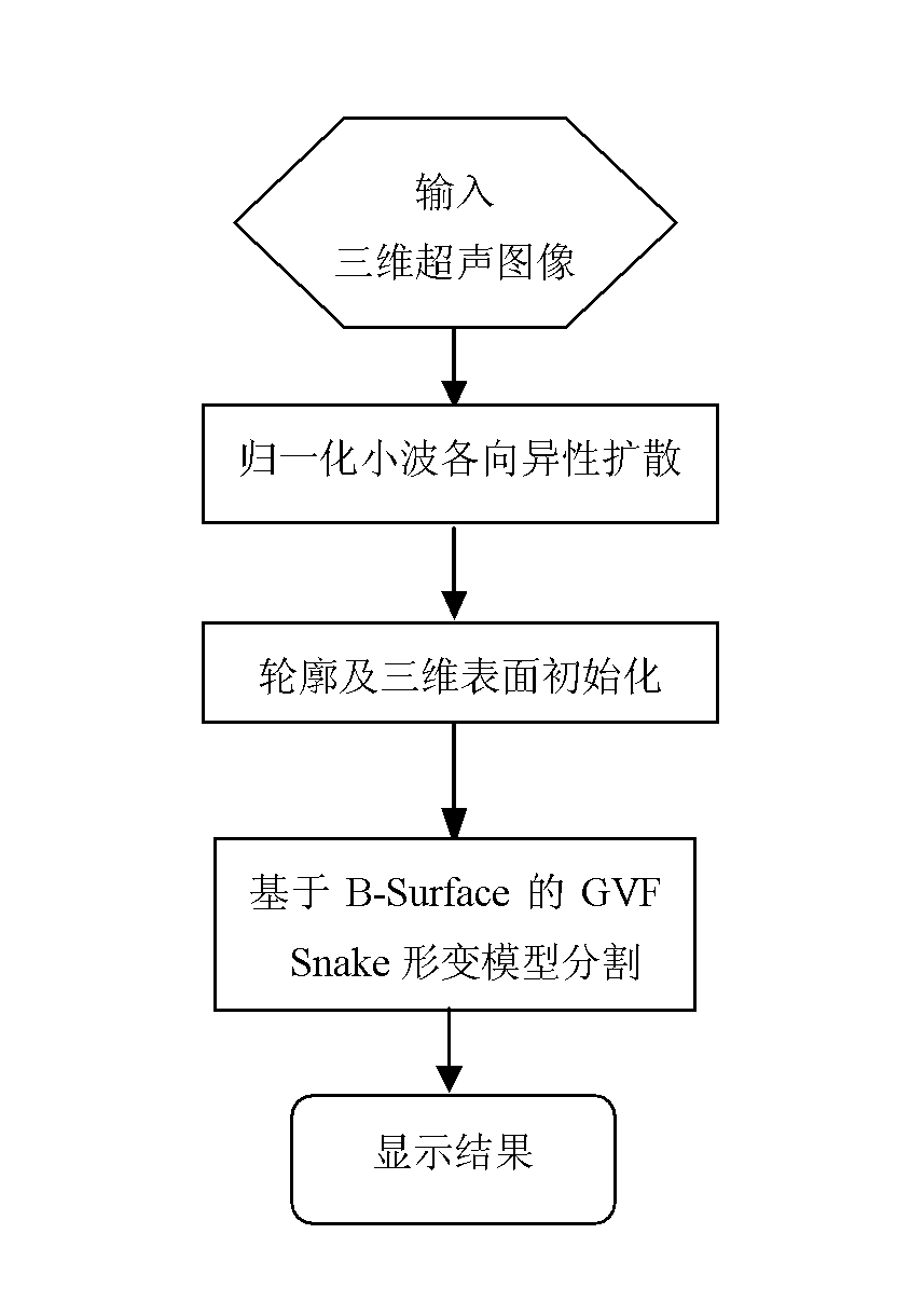
[0065] like figure 1 , 2 Shown, the present invention comprises the following steps:

[0066] 1) Input the three-dimensional ultrasound image to be processed;

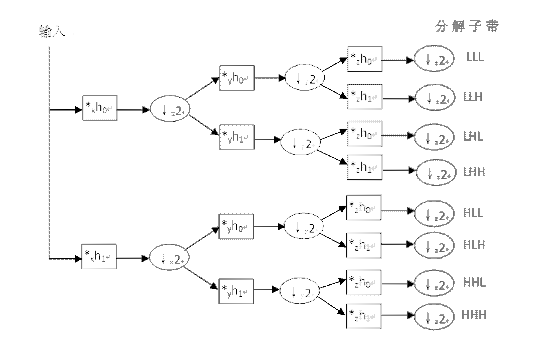
[0067] 2) Carry out three-dimensional wavelet decomposition to the image;

[0068] Decompose the 3D ultrasound image into image 3 Shown are HLL, HLH, HHL, HHH; LHH, LHL, LLH, LLL 8 parts, where LLL is the low frequency part, and the others are high frequency parts.

[0069] 3) Apply normalized anisotropic diffusion to process the high-frequency part to suppress speckle noise;

[0070] The normalized anisotropic diffusion is used to smooth the high-frequency part, that is, the sub-bands HLL, HLH, HHL, HHH; LHH, LHL, LLH 7 parts.

[0071] The speckle multiplicative noise model that the prese...


PUM


Abstract

The invention discloses a method for segmenting a three-dimensional ultrasonic image, belonging to the technical field of the digital image processing. The method comprises the following steps: (1) preprocessing spots of the three-dimensional ultrasonic image by adopting a normalized anisotropic diffusion method of a three-dimensional wavelet according to the characteristics of the three-dimensional ultrasonic image to remove spot noise; (2) initializing the preprocessed three-dimensional ultrasonic image by adopting a Canny edge detection operator; and (3) segmenting the three-dimensional ultrasonic image three-dimensionally by using a B-Surface and GVF Snake based three-dimensional deformation model. The method disclosed by the invention can be used to rapidly and accurately segment the three-dimensional ultrasonic image and particularly has strong noise robustness. The method for automatically segmenting the three-dimensional ultrasonic image can be also used for segmenting other three-dimensional images such as CT (computed tomography) images, MRI (magnetic resonance images) and PET (position-emission tomography) images, thereby having high application value.

Description

technical field [0001] The invention belongs to the field of computer digital image processing, in particular to a segmentation of three-dimensional ultrasonic images. Background technique [0002] Ultrasound imaging is a technology that uses ultrasonic waves of a specific wavelength to generate backscattered signals after the detection body. It has the advantages of high safety, fast imaging speed, convenient operation, and low cost. It is widely used in clinical practice, especially It is the exploration of abdominal organs, such as the heart, liver, kidney, etc., as well as the three-dimensional imaging and real-time four-dimensional dynamic imaging of the fetus. It is one of the most important means of general survey and clinical diagnosis. In addition, three-dimensional ultrasonic imaging technology is also widely used in the monitoring of animal reproduction and development, such as early pregnancy detection, lesion screening, and dynamic evaluation of embryonic growth...

Claims


Application Information

Patent Timeline
IPC IPC(8): G06T7/00G06T5/00
Inventor 郭圣文
Owner SOUTH CHINA UNIV OF TECH
Why Eureka
Try Eureka