
Method for detecting heparins

A detection method and heparin technology, which are used in measurement devices, material analysis by electromagnetic means, instruments, etc., can solve the problems of slow monitoring by fluorescence method, inability to monitor online, affecting accuracy, etc., to shorten the production cycle and shorten the detection. Time, easy operation effect

Active Publication Date: 2012-05-09
YANTAI INST OF COASTAL ZONE RES CHINESE ACAD OF SCI
View PDF5 Cites 14 Cited by
  • Summary
  • Abstract
  • Description
  • Claims
  • Application Information

AI Technical Summary

Problems solved by technology

However, the whole blood activated coagulation time method needs to be measured with whole blood, and the presence of platelets will affect the experimental results; molecular spectroscopy is difficult to achieve online, in situ, real-time, multi-component simultaneous determination, and the detection sensitivity of this method is relatively low. Low; the fluorescence method has defects such as slow monitoring speed, poor continuity, high analysis cost, secondary pollution, and inability to monitor online; high-performance liquid chromatography has high technical requirements for researchers, and complex components in blood will affect the determination accuracy of results

Method used


Image

  • Method for detecting heparins
  • Method for detecting heparins
  • Method for detecting heparins

Examples


Embodiment 1

[0027] Take heparin in the buffer solution tested by this electrode as an example. Its determination steps are as follows:

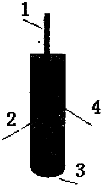
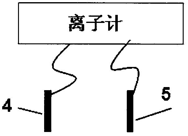
[0028] a. Use ion selective electrode as working electrode, Ag / AgCl (3M KCl) electrode as reference electrode, PXSJ-216L ion meter to measure potential value, ion selective electrode, Ag / AgCl (3M KCl) and PXSJ-216L ion connection (see figure 2 ). Insert the unactivated ion-selective electrode directly into the measuring cell filled with buffer solution, and record the initial potential. The ion selective electrode (see figure 1 ) is inserted with an Ag / AgCl internal reference electrode, and at the same time, 50mM Tris-HCl (pH=7.4) buffer solution and 0.12M NaCl mixture are injected into the ion-selective electrode as the filling solution, and the polymer sensitive film is adhered to the bottom of the electrode.

[0029] Electrode preparation process: get 200mg polymer membrane material, including 0.5wt% protamine, 3wt% tetrakis(dodecyl)-tetrakis(4-c...

Embodiment 2

[0034] First, take 0.12M NaCl solution and configure two spiked samples, the concentrations are 0.05U / ml and 0.2U / ml respectively, measure the initial value of the potential according to Example 1, calculate the initial rate of change of the potential according to the initial value of the potential, and compare Standard working curve (such as Figure 5 ) to calculate the corresponding concentration.

Embodiment 3

[0035] Example 3 The electrode is used to test the heparin in sheep blood. Its determination steps are as follows:

[0036] a. Add sodium citrate to fresh sheep blood to prevent it from coagulating, and use this blood sample as the background electrolyte to prepare heparin samples of different concentrations with known concentrations,

[0037] b. Use the ion-selective electrode as the working electrode, the Ag / AgCl (3M KCl) electrode as the reference electrode, and measure the potential value with the PXSJ-216L ion meter. Ion selective electrode, Ag / AgCl (3M KCl) is connected with PXSJ-216L ion meter (see figure 2 ). Insert the unactivated ion-selective electrode directly into the measuring pool filled with sheep blood, record the initial potential, calculate the initial change rate of the potential according to the initial value of the potential, and use it as a control signal, refer to the standard working curve for the control signal, namely The content of heparin in th...


PUM


Abstract

The invention relates to detection of heparins, in particular to a quick, accurate and sensitive method for detecting heparins. The method comprises the following steps of: testing potential changes caused by adding of heparins of different concentrations by using a potential tester according to specific combination of protamine in a polymer sensitive film phase and heparins in a water phase; drawing a standard work curve according to an initial potential change rate; and obtaining the concentration of heparins in an unknown sample in reference to the standard work curve. In the invention, protamine is added into a polymer film, so that the protamine is not required to be added manually; and an electrode can be directly applied to sample detection without activating, so that the method has the advantages of easiness and convenience for operating, short detection time, low operating cost, suitability for field detection, and the like.

Description

technical field [0001] The invention relates to the detection of heparin, in particular to a fast, accurate and sensitive method for detecting heparin. Background technique [0002] At present, the analytical methods for heparin detection mainly include whole blood activated clotting time method, molecular photometry, fluorescence method and high performance liquid chromatography. However, the whole blood activated coagulation time method needs to be measured with whole blood, and the presence of platelets will affect the experimental results; molecular spectroscopy is difficult to achieve online, in situ, real-time, multi-component simultaneous determination, and the detection sensitivity of this method is relatively low. Low; the fluorescence method has defects such as slow monitoring speed, poor continuity, high analysis cost, secondary pollution, and inability to monitor online; high-performance liquid chromatography has high technical requirements for researchers, and c...

Claims


Application Information

Patent Timeline
IPC IPC(8): G01N27/333
Inventor 秦伟陈燕
Owner YANTAI INST OF COASTAL ZONE RES CHINESE ACAD OF SCI
Learn More