Gene application in inhibition and apoptosis of glioma cell

A glioma cell and gene technology, applied in the application field of gene inhibition and apoptosis in glioma cells, can solve problems such as inability to cure, large brain damage, and easy recurrence

Active Publication Date: 2014-04-02
SHANGHAI UNIV
View PDF4 Cites 0 Cited by
  • Summary
  • Abstract
  • Description
  • Claims
  • Application Information

AI Technical Summary

Problems solved by technology

However, these four treatment methods are very damaging to the brain, easy to relapse, and consume a lot of the patient's body, and they cannot achieve a radical cure.

Method used


Image

  • Gene application in inhibition and apoptosis of glioma cell
  • Gene application in inhibition and apoptosis of glioma cell
  • Gene application in inhibition and apoptosis of glioma cell

Examples


Embodiment 1

[0018] Embodiment one: dcf1 Gene amplification and enzyme digestion identification of recombinant plasmid pEGFP-N2-DCF1

[0019] 1. Using the cDNA of HeLa cells as a template for PCR amplification, in dcf1 EcoRI and BamHI restriction sites were introduced at both ends of the primers respectively. The upstream primer is (5'-CGGAATTCATGGCGGCGCCGAAGGGGAG-3'),

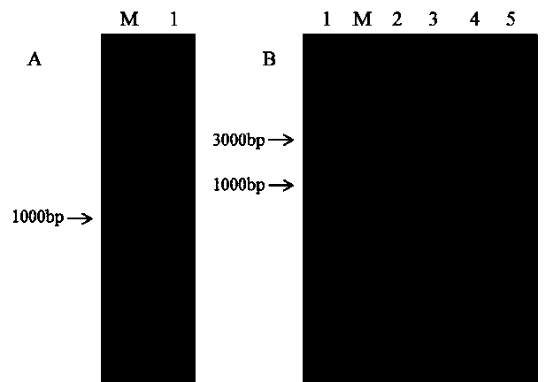
[0020] The downstream primer is (5'-CGGGATCCGTAAAATTTCAGAATGAGCA-3'), Tm is 53 degrees Celsius, and after 30 cycles, the target fragment of 972bp is obtained ( figure 1 A).

[0021]2. After the positive recombinant plasmid pEGFP-N2-DCF1 was obtained, the recombinant was identified by double enzyme digestion. Two enzymes, EcoRI and BamHI, were added to the enzyme digestion system at the same time. As a control, pEGFP-N2 EcoRI single enzyme digestion was used, and the reaction was performed at 37 degrees Celsius for 2 hours. The enzyme digestion results were identified by electrophoresis, and a band was released at 97...

Embodiment 2

[0022] Example 2: Transfection of plasmid pEGFP-N2-DCF1

[0023] After the U251 cell line was plated in a 24-well plate for 24 hours, the plasmid pEGFP-N2-DCF1 was transfected by Lipofectamine® 2000 Transfection Reagent cationic liposome transfection method. 1ul lipo2000 per well, 1ug ​​plasmid pEGFP-

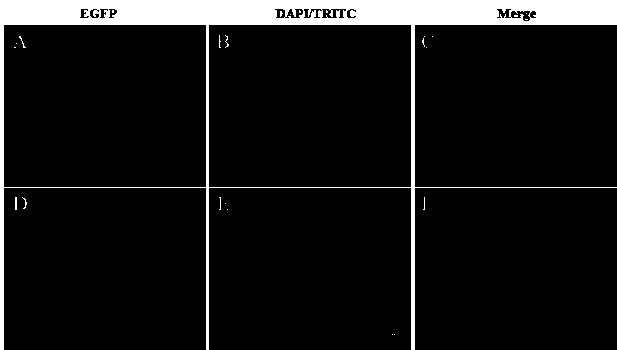
[0024] N2-DCF1 was dissolved in 50ul serum-free DMEM medium respectively, and after standing at room temperature for 5 minutes, the two were mixed evenly, and standing at room temperature for 20 minutes. The medium in the well plate was replaced with 400ul serum-free DMEM, and 100ul of the above mixed product was added. After culturing at 37°C for 6 hours, the medium was replaced with DMEM containing 10% fetal bovine serum. Fluorescence was detected 48 hours after transfection ( figure 2 A-C).

Embodiment 3

[0025] Example 3: Cell Immunofluorescence

[0026] Forty-eight hours after transfection of U251 cells, discard the medium and wash three times with PBS. Fix with 4% PFA for 10 minutes and wash with PBS three times. Permeabilize with 1‰Triton-X100 for 10 minutes, wash with PBS three times. Block with 2% BSA-PBS for 1 hour at room temperature. Dilute DCF1 rabbit polyclonal antibody (1:700) with 1% BSA-PBS, incubate at 37°C for 1 hour, wash with PBS three times. Dilute TRITC fluorescent secondary antibody (1:300) with 1% BSA-PBS, incubate at 37°C for 40 minutes in the dark, wash with PBS three times, and observe under a fluorescence microscope ( figure 2 D-F).


PUM


Abstract

The present invention relates the application of a human source dcf1 gene in the inhibition of U251 glioma cell line proliferation and in the induction of U251 glioma cell line apoptosis. The present invention makes judgments for dcf1-induced U251 cell line apoptosis by the methods of immune electron microscope, atomic force microscope, CCK8 proliferation detection, JC-1 staining, and nude mouce tumor heterotopic transplantation. The results show that: the dcf1 causes mitochondrial structure lesion and membrane potential reduction by locating in mitochondria, thereby causing apoptosis-related gene expression change, and eventually leading to apoptosis. The nude mouce experiments show that the dcf1 can significantly reduce glioma tumor volume and inhibit tumor growth.

Description

technical field [0001] The present invention relates to a human source dcf1 new uses. especially human source dcf1 Application of gene in inhibition and apoptosis of glioma cells. Background technique [0002] Glioma is a tumor arising from glial cells (astrocytes, oligodendrocytes, ependyma, etc.) differentiated from the neuroectoderm. Glioma ranks first in the incidence of intracranial tumors, accounting for about 45%. The treatment of glioma is a difficult problem in neurosurgery. Due to its high mortality rate, poor prognosis, uncontrollable cell proliferation, slow cell apoptosis, and strong invasion of tumor cells, the treatment of glioma faces a huge challenge. The average survival period of surgery plus radiotherapy and chemotherapy after illness is only 8 to 11 months. At present, there are mainly the following methods for tumor treatment: surgical treatment, chemical drugs, therapeutic radiation therapy, and photodynamic therapy. But these four kinds of treat...

Claims


Application Information

Patent Timeline
Patent Type & Authority Patents(China)
IPC IPC(8): A61K48/00A61P35/00
Inventor 文铁桥谢雨琼杨眉杨清波
Owner SHANGHAI UNIV
Why Eureka
Try Eureka