Method for detecting phytase

A detection method and technology for phytase, applied in the preparation of test samples, measurement of color/spectral characteristics, etc., can solve the problems affecting the accurate quantification of phytase activity, background value interference, etc., to avoid background value interference, fast measured effect

CN102980854AInactive Publication Date: 2013-03-20NANJING AGRICULTURAL UNIVERSITY
1 Cites 5 Cited by

Patent Information

Authority / Receiving Office
CN · China
Patent Type
Applications(China)
Current Assignee / Owner
Publication Date
2013-03-20
Estimated Expiration
Not applicable · inactive patent

Smart Images

  • Figure 1
    Figure 1
  • Figure 2
    Figure 2
  • Figure 3
    Figure 3
Patent Text Reader

Abstract

The invention belongs to the field of biological detection, and relates to a method for detecting phytase in samples. The method mainly comprises the following steps of: forming a surface rapid reaction system by combining phytase with phytic acid in a solvent phase by coupling dispersing-type nano microspheres and phytic acid molecules by utilizing a carboxyl-amino chemical coupling method, wherein a product orthophosphate enters the solvent phase directly, and unreacted inositol derivatives are still remained in solid-phase microspheres; removing the microspheres through low-temperature and high-speed centrifuging, adding color developing agents into the solvent phase under the acidic condition so as to generate a blue compound, and carrying out colorimetric determination at the wavelength of 700nm. For the method, the phytase activity is determined rapidly, meanwhile, the unreacted inositol derivatives are still remained in solid-phase microspheres, and the background value interference caused by the reaction of the incompletely reacted phytic acid molecules and the color developing agents in the conventional method is prevented, so that the trace amount determination becomes possible; and the method for detecting phytase is suitable for trace amount phytase sample determination for specific environment samples, such as soil, water bodies and rhizosphere residues.
Need to check novelty before this filing date? Find Prior Art

Description

technical field

[0001] The invention belongs to the field of biological detection and relates to a method for detecting phytase in a sample. Background technique

[0002] Phytase widely exists in plants, animal tissues and microorganisms. Phytases are divided into 3 classes: phytate-3-phosphate hydrolases, phytate-6-phosphate hydrolases and nonspecific orthophosphate monoester phosphohydrolases. There are three sources of phytase found at present: one is the natural phytase in plants, such as the natural phytase contained in grain grains such as wheat, barley, rye and their processing by-products; the other is microbial source The phytase is the phytase that is currently considered to have the most potential; the third is a phytase-producing bacterium isolated from the bovine rumen, and can also produce other enzymes with nutritional functions.

[0003] Phytase can catalyze the hydrolysis of phytic acid and its salts into inositol and phosphoric acid (salt), has a special ...

Examples

Embodiment 1

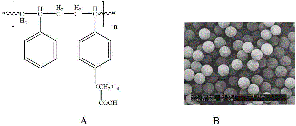
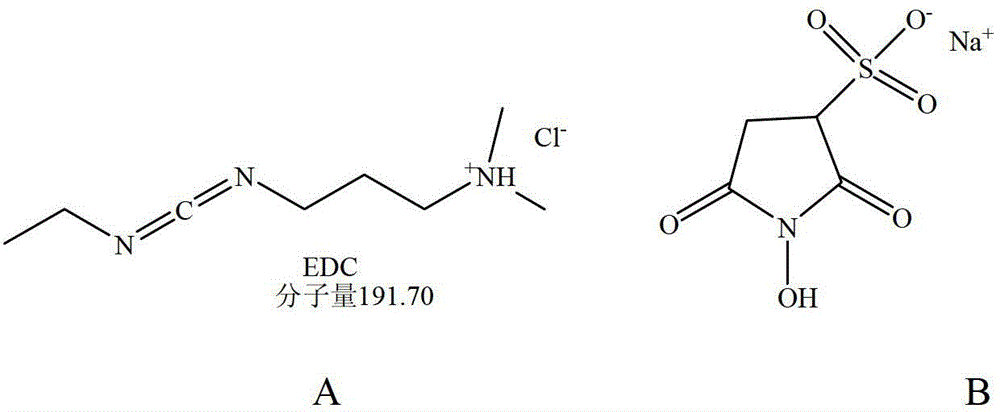
[0020] like figure 1 As shown, the nano-microspheres are monodisperse carboxylated polystyrene microspheres with a particle diameter of figure 2 A) Activation for 30min, then add 1ml 50mM protective agent Sulfo-NHS (N-hydroxysulfosuccinimide) ( figure 2 B) 15 minutes to make the microspheres form a stable active ester intermediate, then add 2ml of 5mM sodium phytate and incubate at 30°C for 1-2 hours, collect the nanospheres coupled with phytic acid molecules after washing.

Embodiment 2

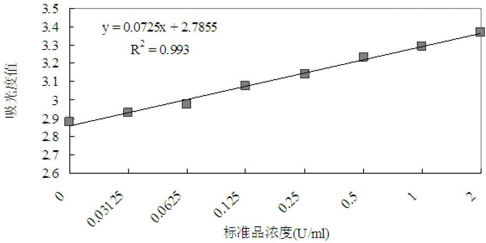
[0022] As shown in Table 1, the nano-microspheres coupled with phytic acid molecules were prepared into a 2.5% (w / v%, weight / volume percentage, specifically g / 100ml) suspension with 0.1M acetic acid buffer, and the pH was adjusted to 5. 0. Take 80 μl of microspheres and add 20 μl of sample or phytase standard. Using phytase pure protein as a standard, set up 8 concentration gradients (2U / ml, 1U / ml, 0.5U / ml, 0.25U / ml, 0.125U / ml, 0.0625U / ml, 0.03125U / ml and 0U / ml). After preheating at 37°C for 5 minutes, mix well for 5 minutes, and keep at 37°C for 30 minutes. Centrifuge at 4°C and 15,000×g for 20 min, and collect the supernatant. Next, add 100 μl of 5% (w / v%, specifically g / 100ml) trichloroacetic acid (stop solution) and 100 μl of chromogen (1.5% (w / v%, specifically g / 100ml) sodium molybdate and 2.7% (w / v%, specifically refers to g / 100ml) ferrous sulfate according to the ratio of 4:1, ready to use), immediately after mixing, colorimetric measurement at a wavelength of 700n...