Ultrasonic training system based on CT (Computed Tomography) image simulation and positioning

A CT imaging and training system technology, applied in the field of medical ultrasound training, can solve problems such as difficulty in meeting the real-time requirements of medical ultrasound training, incomplete three-dimensional structure information of images, blurred display effect, etc. Improve applicability and practicality, facilitate the effect of ultrasonic simulation

Active Publication Date: 2013-09-11
ARIEMEDI MEDICAL SCI BEIJING CO LTD
View PDF5 Cites 20 Cited by
  • Summary
  • Abstract
  • Description
  • Claims
  • Application Information

AI Technical Summary

Problems solved by technology

[0005] 1. The computational complexity of the ultrasound image simulation method based on CT volume data is high, and it is difficult to meet the real-time requirements of medical ultrasound training;
[0006] 2. Since the accuracy of 3D volume data rendering and image fusion display is directly proportional to the complexity of the algorithm, when real-time performance is met, the 3D structure information of the image is incomplete and the display effect is blurred;

Method used


Image

  • Ultrasonic training system based on CT (Computed Tomography) image simulation and positioning
  • Ultrasonic training system based on CT (Computed Tomography) image simulation and positioning
  • Ultrasonic training system based on CT (Computed Tomography) image simulation and positioning

Examples


Embodiment Construction

[0034] The present invention will be described in detail below in conjunction with specific embodiments and drawings, but the present invention is not limited thereto.

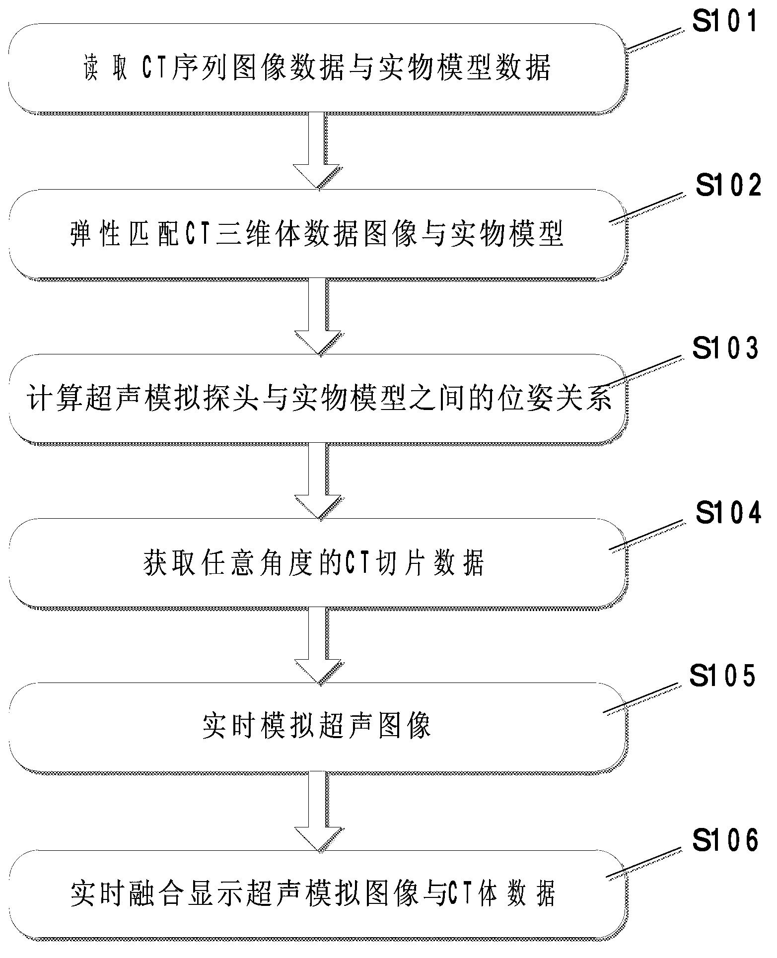
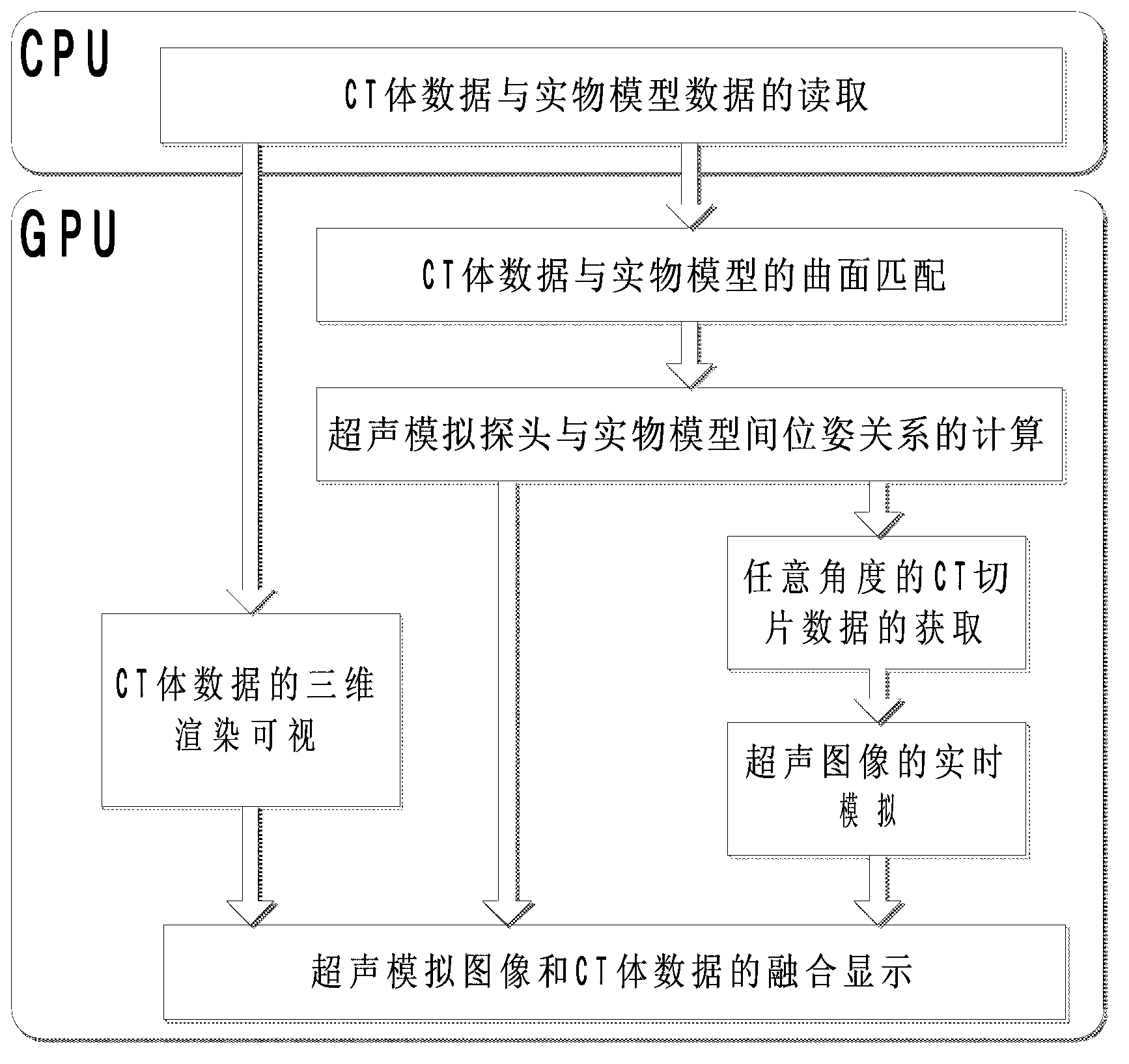
[0035] attached figure 1 To reconstruct the flow chart, the ultrasound simulation training system includes the following steps:

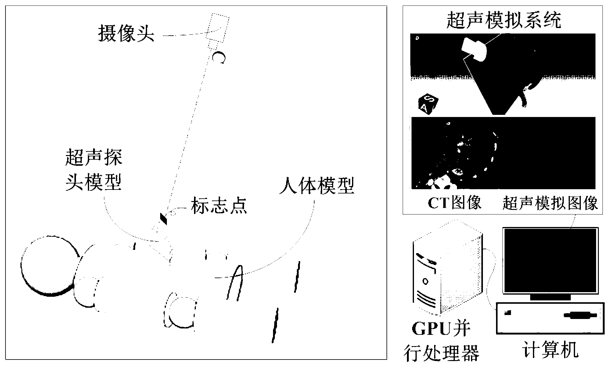
[0036] Step S101, read the CT sequence image data as the source image of the ultrasound simulation, and read the data of the solid model of the human abdominal cavity.

[0037] Step S102, using the read human abdominal cavity solid model data as a standard and the read CT sequence image data as an image to be registered, using an octree-based matching algorithm to complete surface matching between the CT volume data and the human abdominal cavity model data. The process of elastic registration based on octree algorithm is as follows:

[0038] (1) In the image to be registered, that is, select a certain number of marker points on the data surface of the human abdominal cavity model...


PUM


Abstract

The invention provides an ultrasonic training system based on CT (Computed Tomography) image simulation and positioning. Ultrasound image simulation and CT volumetric data rendering are accelerated to be achieved through a GPU (Graphics Processing Unit) and the real-time performance of the system is improved. A curved surface matching module is used for performing surface matching on read human body CT volumetric data and physical model data with a physical model as the standard and achieving elastic transformation of a curved surface based on an interpolation method of thin plate splines; an ultrasonic simulation probe position tracking module is used for performing real-time calculation on ultrasonic simulation probe positions relative to the physical model by a marking point tracking method and obtaining arbitrary angle CT slice images according to a position matrix; an image enhancement and ultrasonic image simulation generation module is used for improving the vessel contrast ratio in CT images by a multi-scale enhancement method and achieving the ultrasound image simulation based on the CT volumetric data; and an integration display module is used for accelerating to achieve rendering display of the CT volumetric data based on CUDA (Compute Unified Device Architecture) and integrating and displaying ultrasound simulation images and three-dimensional CT images according to the obtained position matrix.

Description

technical field [0001] The invention relates to an ultrasound training system based on CT image simulation and positioning, which is suitable for the field of medical ultrasound training. Background technique [0002] When ultrasound propagates in the human body, due to the differences in the acoustic properties of various tissues in the human body, the ultrasound will produce physical phenomena such as reflection, refraction, scattering, and Doppler frequency shift at the interface between two different tissues. The application of ultrasonic diagnostic equipment to receive these reflected and scattered signals can display various tissues and the morphology of their lesions. Combined with pathology and clinical medicine, physicians can make accurate diagnoses on the location, nature, and degree of dysfunction of lesions. [0003] In addition, because ultrasound has no radiation and fast imaging speed, it can be widely used in the guidance process of clinical minimally invasi...

Claims


Application Information

Patent Timeline
Patent Type & Authority Applications(China)
IPC IPC(8): G09B23/28
CPCG09B23/286G09B23/30
Inventor 杨健王涌天刘越丛伟建宋凌
Owner ARIEMEDI MEDICAL SCI BEIJING CO LTD
Why Eureka
Try Eureka