
A sample preparation kit for detecting molecules on the membrane surface of pig peripheral blood and spleen lymphocytes and its preparation method

A sample preparation and peripheral blood technology, applied in the preparation of test samples, particle and sedimentation analysis, measuring devices, etc., to overcome bottlenecks, flow cytometry detection results are efficient and stable, and inhibit non-specific binding effects

Active Publication Date: 2018-03-13
昆明云中美农牧科技有限公司 +2
View PDF5 Cites 0 Cited by
  • Summary
  • Abstract
  • Description
  • Claims
  • Application Information

AI Technical Summary

Problems solved by technology

However, so far, there is no mature and feasible method for sample preparation suitable for flow cytometry detection in grass-roots pig farms

Method used


Image

  • A sample preparation kit for detecting molecules on the membrane surface of pig peripheral blood and spleen lymphocytes and its preparation method
  • A sample preparation kit for detecting molecules on the membrane surface of pig peripheral blood and spleen lymphocytes and its preparation method

Examples


Embodiment 1

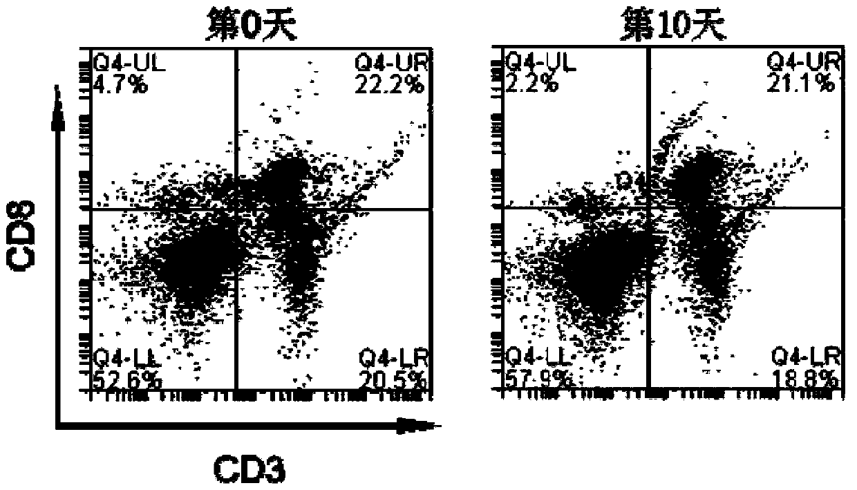
[0034] The method of the invention is used to prepare samples for detection of CD3 and CD8 molecule expression in peripheral blood lymphocytes of porcine.

[0035] The kit used includes buffer I, buffer II and buffer III, the buffer I is: PBS buffer containing 500 U / ml penicillin, 500 μg / ml streptomycin, 5 μg / ml amphotericin B, pH 7.4; the buffer II is containing 2mMEDTA, 0.5% BSA, 500U / ml penicillin, 500 μg / ml streptomycin, the PBS buffer of 5 μg / ml amphotericin B, the pH value is 7.4; the buffer III with 0.1% NaN 3 , 2% paraformaldehyde in PBS buffer, pH 7.4.

[0036] This embodiment also uses erythrocyte lysate, the main component is: 0.16M NH 4 Cl, 0.13mM EDTA, 12mM NaHCO 3 , pH value is 7.2.

[0037] The specific steps are:

[0038]1) Collect 2ml porcine anterior vena cava blood with EDTA anticoagulant vacuum blood collection tube, and transfer to 15ml centrifuge tube;

[0039] 2) Add 6ml of erythrocyte lysate, invert and mix;

[0040] 3) Incubate on ice for 5 minu...

Embodiment 2

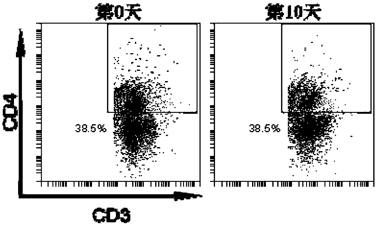
[0051] The method of the invention is used to prepare samples for detection of CD3 and CD4 molecule expression in porcine spleen lymphocytes.

[0052] The kit used includes buffer I, buffer II and buffer III, the buffer I is: PBS buffer containing 100 U / ml penicillin, 800 μg / ml streptomycin, 5 μg / ml amphotericin B, pH 7.4; the buffer II is containing 2mMEDTA, 0.4% BSA, 300U / ml penicillin, 600 μg / ml streptomycin, the PBS buffer of 7 μg / ml amphotericin B, the pH value is 7.4; the buffer III with 0.1% NaN 3 , 5% paraformaldehyde in PBS buffer, pH 7.4.

[0053] This embodiment also uses erythrocyte lysate, the main component is: 0.16M NH 4 Cl, 0.13mM EDTA, 12mM NaHCO 3 , pH value is 7.2.

[0054] The specific steps are:

[0055] 1) Cut about 0.5cm of spleen tissue 3 Put it in a plate, remove the capsule and connective tissue, grind the spleen on a 200-mesh nylon membrane, suspend the ground cells in 2ml buffer I through the nylon mesh, and transfer the cell suspension to a 1...


PUM


Abstract

The invention discloses a sample preparation kit suitable for flow cytometry detection of molecules on the membrane surface of porcine peripheral blood and spleen lymphocytes and a preparation method thereof. Wherein, in the preparation method, the biological sample is obtained from the peripheral blood or spleen of the pig and made into a crude cell suspension; after the crude extract is transferred to a centrifuge tube, the red blood cell lysate is added to lyse the red blood cells in an ice bath, and the supernatant is removed by centrifugation. Collect the precipitated cells; resuspend the cells with buffer I, and count the cells; resuspend the cells with buffer II, add fluorescein-labeled cell membrane surface antibody, and ice-bath for 30min; wash the cells with buffer I, and Cells were suspended in buffer III. The samples prepared by the present invention can be stored at room temperature or transported over long distances for detection by a flow cytometer, and the detection results are efficient and stable.

Description

technical field [0001] The invention relates to a flow cytometric detection technology for molecules on the membrane surface of pig peripheral blood and spleen lymphocytes, and in particular provides a flow cytometric sample preparation method suitable for low-level pig farm laboratories with poor conditions. Background technique [0002] my country is a big country in pork production and consumption. Internationally, my country's pig stock, slaughter volume and pork output have ranked first in the world for many years. According to the National Bureau of Statistics, my country's pork output was 53.35 million tons in 2012, a year-on-year increase of 5.6%. my country's aquaculture industry plays a pivotal role in the world. Since the 1980s, China's pig raising industry has entered a period of transition from farmers raising pigs to intensification and industrialization, and the science and technology of pig raising has also made great progress. However, the pig industry is...

Claims


Application Information

Patent Timeline
Patent Type & Authority Patents(China)
IPC IPC(8): G01N1/28G01N15/14
Inventor 苏华荔王安如
Owner 昆明云中美农牧科技有限公司
Learn More