Vaginal secretion analysis system

A vaginal secretion and analysis system technology, applied in the field of vaginal secretion analysis system, can solve problems such as poor reliability of test results of trichomoniasis and fungi, unsystematic results, unfavorable clinical treatment, etc., so as to improve clinical diagnosis and treatment efficiency and rapid analysis report. , to avoid the effect of subjectivity

Active Publication Date: 2015-03-25
北京中生金域诊断技术股份有限公司
View PDF7 Cites 0 Cited by
  • Summary
  • Abstract
  • Description
  • Claims
  • Application Information

AI Technical Summary

Problems solved by technology

[0004] The first type is the morphological wet film examination method using a microscope, which can directly observe the formed components of vaginal secretions, and detect normal bacteria, trichomonas, fungi and other miscellaneous bacteria, epithelial cells, and white blood cells in vaginal secretions. , but the antibacterial function of normal dominant bacteria cannot be detected, the pathogenicity of miscellaneous bacteria, aerobic bacteria or anaerobic bacteria cannot be distinguished, and the pathogenicity of fungi cannot be distinguished, etc.
[0005] The other is to use biochemical methods to conduct dry chemical detection of vaginal secretions, mainly through the detection of indicators such as preformed enzymes and metabolites of normal dominant bacteria and pathogenic bacteria, which can reflect the functionality of normal vaginal bacteria and pathogenic bacteria However, this method has poor reliability and poor specificity for trichomonas and fungus test results, and cannot reflect the cleanliness. Moreover, due to the strong subjectivity and heavy workload of manual interpretation, the detection speed will be affected
[0006] At present, clinically, vaginal secretions are mostly tested in one way. The results of the test are not systematic and comprehensive, which may easily lead to misdiagnosis and missed diagnosis, which is not conducive to clinical treatment.
[0007] In summary, the existing vaginal secretion detection system has problems such as low detection efficiency and poor reliability of detection results.

Method used


Image

  • Vaginal secretion analysis system
  • Vaginal secretion analysis system
  • Vaginal secretion analysis system

Examples


Embodiment 1

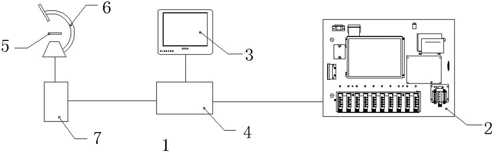
[0035] Embodiment 1: as figure 1 , 4 As shown, the vaginal secretion analysis system of the present invention includes:

[0036] The morphology detection unit 1 is used to detect the formed components of vaginal secretions;

[0037] Biochemical detection unit 2, used to detect preformed enzymes and metabolites in vaginal secretions;

[0038] Human-computer interaction device 3, for data interaction with data processing unit 4;

[0039] The data processing unit 4 is used to process the data acquired by the physical detection unit 1 , the biochemical detection unit 2 and the human-computer interaction device 3 to obtain the analysis result of vaginal secretions.

[0040] The physical detection unit includes: a stage 5 , a microscope 6 and a CCD 7 , and the CCD 7 is used to transmit the collected images to the data processing unit 4 .

[0041] The biochemical detection unit 2 includes:

[0042] The color detection module 8 is used to realize the color measurement of the samp...

Embodiment 2

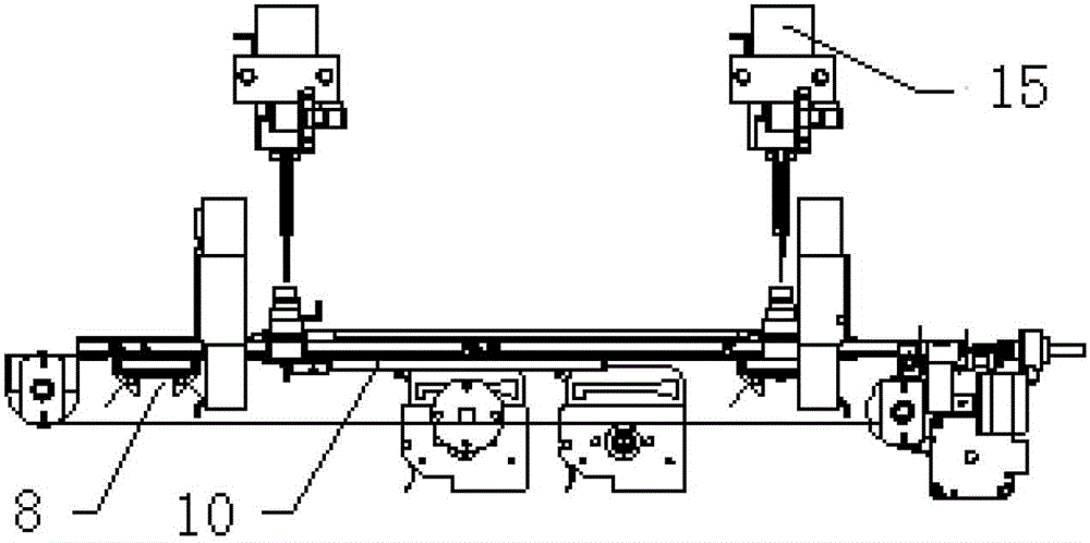
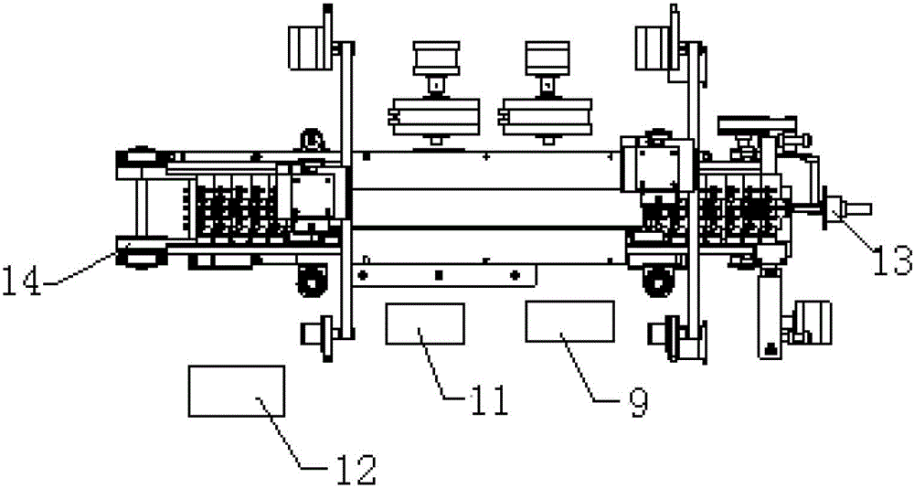
[0051] Embodiment 2: as Figure 1-3 As shown, the difference between this embodiment and Embodiment 1 is that the biochemical detection unit 2 in this embodiment also includes: a feeding mechanism 13 for realizing the automatic feeding of the sample to be tested, and a delivery mechanism 13 for transmitting the sample to be tested. Mechanism 14 and a sample loading arm mechanism 15 used for automatic quantitative sample loading during testing.

[0052] The working process of biochemical detection unit 2 in the present embodiment:

[0053] After scanning the bar code information of the sample to be tested with the external scanning gun 12, input relevant information on the touch screen 9, after the input is completed, put the sample to be tested into the entrance of the feeding mechanism 13, and the photocoupler at the entrance detects After receiving the sample arrival signal, the feeding mechanism 13 automatically feeds the material. After the feeding is completed, the trans...

Embodiment 3

[0057] Embodiment 3: A method for analyzing vaginal secretions using the aforementioned system, comprising the following steps:

[0058]The morphology detection unit detects the color, character and smell of vaginal secretions, the shape and quantity of normal bacteria, epithelial cells, miscellaneous bacteria, pathogenic microorganisms, and white blood cells or pus cells;

[0059] The biochemical detection unit detects the biochemical functional characteristics of normal bacteria, miscellaneous bacteria and pathogenic microorganisms in vaginal secretions;

[0060] The data processing unit comprehensively analyzes the detection results of the morphological detection unit and the biochemical detection unit, as well as the information input by the human-computer interaction device to obtain the vaginal infection situation.

[0061] The information on the formed components of vaginal secretions includes: physical indicators such as the color, character, and smell of vaginal secre...


PUM


Abstract

The invention discloses a vaginal secretion analysis system. The system includes a morphological detection unit for detecting formed elements of vaginal secretions, a biochemical detection unit for biochemical detection of the vaginal secretions, a human-computer interaction device for data interaction with a data processing unit, and the data processing unit for processing data obtained through the morphological detection unit, the biochemical detection unit and the human-computer interaction device to obtain analysis results of the vaginal secretions. The vaginal secretions are subjected to a morphological microscopic examination, and synchronous detection and synchronous analysis of pre-produced enzyme spectra and metabolite spectra of normal bacteria and pathogenic bacteria, so that morphological results and biochemical results are combined and supplemented with each other, and thus misdiagnosis, such as a missed diagnosis, caused by no completeness, no comprehension and no systematicness of only consideration of the physics or biochemical test results is avoided. The vaginal secretion analysis system can accurately evaluate the vaginal micro ecology, and finds out all common vaginitis and vaginal infection pathogenic microorganisms.

Description

technical field [0001] The invention relates to the technical field of medical clinical testing, in particular to a vaginal secretion analysis system. Background technique [0002] At present, the prevention and treatment of vaginal infection clinically faces the problems of low cure rate and high recurrence rate. The main reason for this phenomenon is that clinicians are often eager to treat without first identifying the pathogenic microorganism, which is the most common cause of treatment failure. [0003] Routine detection of vaginal discharge (referred to as leucorrhea routine) is an important medical examination item that is widely used clinically. The methods for routine detection of vaginal discharge in clinical practice are divided into two categories: [0004] The first type is the morphological wet film examination method using a microscope, which can directly observe the formed components of vaginal secretions, and detect normal bacteria, trichomonas, fungi and ot...

Claims


Application Information

Patent Timeline
Patent Type & Authority Patents(China)
IPC IPC(8): G01N35/00
Inventor 王加义王淳玉张永威孙滨赵玉林
Owner 北京中生金域诊断技术股份有限公司
Why Eureka
Try Eureka