Airway epithelium-porous silk fibroin protein complex, and preparation method and application thereof

A technology of porous silk and respiratory tract, which is applied to the culture of respiratory tract epithelium on porous silk fibroin, and the application field of respiratory epithelium-porous silk fibroin complex in the reconstruction of tracheal defect to achieve the effect of reducing complications

Inactive Publication Date: 2014-01-15
AFFILIATED HUSN HOSPITAL OF FUDAN UNIV +1
View PDF6 Cites 1 Cited by
  • Summary
  • Abstract
  • Description
  • Claims
  • Application Information

AI Technical Summary

Problems solved by technology

So far, there is no report on the application of silk fibroin in respiratory epithelial culture at home and abroad.

Method used


Image

  • Airway epithelium-porous silk fibroin protein complex, and preparation method and application thereof
  • Airway epithelium-porous silk fibroin protein complex, and preparation method and application thereof
  • Airway epithelium-porous silk fibroin protein complex, and preparation method and application thereof

Examples


Embodiment 1

[0029] Example 1 Preparation of porous silk fibroin

[0030] The waste mulberry silkworm raw silk is degummed, and the degummed silk is dissolved in LiBr aqueous solution, filtered, and concentrated to make a silk fibroin solution with a concentration of about 20%. Add n-butanol at a volume ratio of 2:1, stir at a low speed, pour the mixed solution of silk fibroin and n-butanol into a self-prepared mold, and obtain a white porous scaffold after freeze-drying. The average pore size of the scaffold is 100-200 μm. Trim it to a diameter of 0.5cm, a thickness of 1mm, and a spare size.

Embodiment 2

[0031] Example 2 Primary Culture of Human Respiratory Tract Epithelium

[0032] After lobectomy, the specimens were taken about 1 cm away from the bronchi of the tumor site, soaked in RPMI1640 culture medium containing 10% FBC immediately, and used 1×10 5 U / L penicillin and 1×10 5Wash the bronchial mucosa three times with normal saline with U / L streptomycin, then wash it three times with PBS, place it in a 60cm culture dish, soak it in RIPM1640 culture medium containing 10% FBC, and use a cell brush for endoscopy (AF-1810XB ) Gently scrape each place on the bronchial mucosa about 7-8 times. The surface of the bronchial mucosa was washed with RPMI1640 culture solution, and the culture solution in the culture dish was centrifuged at 1000 rpm for 5 minutes. Discard the supernatant, wash again with RPMI1640 culture solution containing 10% FBC (centrifuge at 1000 rpm × 5 minutes), discard the supernatant, add 3ml RPMI1640 culture solution containing 10% FBC, blow gently with a di...

Embodiment 3

[0033] Example 3 Composite culture of human airway epithelium and porous silk fibroin scaffold

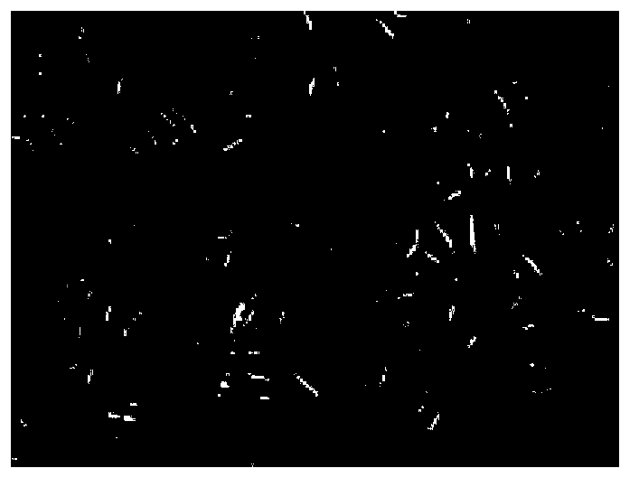
[0034] When the growth of human airway epithelial cells reached 80% confluence, they were digested with trypsin, and the cell concentration was adjusted to 1×10 5 / cm 2 Human airway epithelial cells were inoculated onto porous silk fibroin at a density of 37°C in a 5% CO2 cell incubator for 21 days. Scanning electron microscopy and calcein staining were performed regularly to observe the growth.

[0035] The results showed that respiratory epithelial cells were inoculated on the porous silk fibroin for 3 days, the respiratory epithelial cells adhered well on the silk fibroin scaffold at 3 days, the respiratory epithelium grew in sheets on the silk fibroin material at 14 days, and the respiratory epithelial growth was still active at 21 days; Viable cells were stained with Calcein AM, which confirmed the good viability of the cells.


PUM

PropertyMeasurementUnit
pore sizeaaaaaaaaaa

Abstract

The invention belongs to the fields of tissue engineering and a medical material, relates to an airway epithelium-porous silk fibroin protein complex, and a preparation method and application thereof, and particularly relates to a method for carrying out airway epithelium cultivation on porous silk fibroin protein, and application of the airway epithelium-porous silk fibroin protein complex in trachea defect reconstruction. The porous silk fibroin is adopted as a bracket material; a primary human airway epithelium cell is taken as a seed cell; the airway epithelium cell is subjected to composite cultivation on the porous silk fibroin protein, so as to form the airway epithelium-porous silk fibroin protein complex. The complex is covered on the inner wall of an artificial trachea, so that the inner wall of the artificial trachea is covered by respiratory mucosa, so as to be closer to a normal trachea structure; the artificial trachea can be further applied to preparation of a tissue engineering trachea, repair of clinical tracheal mucosa and clinical tracheal defect reconstruction, and reduction of postoperative complications of tracheal reconstruction as the medical material.

Description

technical field [0001] The invention belongs to the field of tissue engineering and medical materials, and relates to a respiratory epithelium-porous silk fibroin complex and its preparation method and application, especially a method for culturing respiratory epithelium on porous silk fibroin, and respiratory epithelium-porous silk fibroin Application of composites in reconstruction of tracheal defects. Background technique [0002] According to reports, tracheal defects caused by tumors, trauma, and congenital malformations are common in clinical medicine, and when the length of the defect exceeds half of the total length of the trachea (> 6cm), it will not be possible to carry out the operation due to excessive tension of the anastomotic port resulting in luminal leakage. Direct anastomosis requires tracheal reconstruction. The reconstructed trachea in clinical practice must meet the following conditions: sufficient strength and elasticity to maintain patency of the l...

Claims


Application Information

Patent Timeline
Patent Type & Authority Applications(China)
IPC IPC(8): A61L27/38A61L27/22A61L27/54
Inventor 陈仲春赵霞文建川陈新严文洪邵正中
Owner AFFILIATED HUSN HOSPITAL OF FUDAN UNIV
Why Eureka
Try Eureka