Separation method and culture method for umbilical cord mesenchymal stem cells
A separation method and stem cell technology, applied in animal cells, vertebrate cells, bone/connective tissue cells, etc., can solve the problems of insufficient adherence and growth support ability of primary cells, avoid heterologous contamination, and achieve good results. Supporting role, effect of guaranteeing batch stability
- Summary
- Abstract
- Description
- Claims
- Application Information
AI Technical Summary
Problems solved by technology
Method used
Image
Examples
Embodiment 1
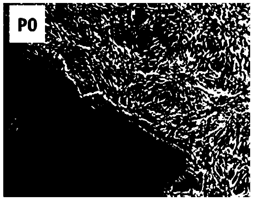
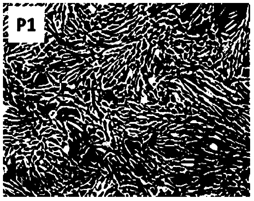
[0067] This embodiment provides a method for isolating and culturing umbilical cord mesenchymal stem cells, which includes the following steps:
[0068] 1. Obtain 15cm of healthy newborn umbilical cord tissue, and wash it with PBS buffer solution containing 2wt% penicillin-streptomycin (final concentration of penicillin 100U / mL, final concentration of streptomycin 0.01g / 100mL) to fully remove blood stains;
[0069] 2. Cut the washed umbilical cord evenly into small sections of 3-4cm in length, perform mechanical separation, bluntly peel off Wharton’s jelly, remove the umbilical artery and umbilical vein at the same time, use ophthalmic scissors to cut the stripped Wharton’s jelly into 1mm 3 -3mm 3 small pieces to obtain Wharton's jelly tissue pieces.
[0070] 3. Take a 10cm petri dish, coat it with 0.2wt% gelatin, and place it at 37°C, 5% CO 2 Incubate for 1 hour in an incubator with no Ca 2+ , Mg 2+ Wash with PBS buffer to remove residual gelatin.
[0071] 4. Resuspend ...
Embodiment 2
[0080] This embodiment provides a method for isolating and culturing umbilical cord mesenchymal stem cells, which includes the following steps:
[0081] 1. Obtain 10cm of umbilical cord tissue from caesarean section neonates, and use PBS buffer solution containing 5wt% penicillin-streptomycin (final concentration of penicillin 100U / mL, final concentration of streptomycin 0.01g / 100mL) to fully remove blood stains;
[0082] 2. Cut the washed umbilical cord evenly into 3-4cm long sections, perform mechanical separation, bluntly peel off Wharton's jelly, and remove the umbilical artery and umbilical vein at the same time;
[0083] 3. Cut the peeled Wharton's glue into 1mm 3 -3mm 3 Add 15 mL of a mixture of collagenase A with a concentration of 1 mg / mL and neutral protease with a concentration of 1 mg / mL (the volume ratio of the two is 1:2), and place at 37 ° C, 5% CO 2 Digested in an incubator for 2 hours, during which time it was taken out every 20 minutes to detect the digesti...
Embodiment 3
[0089] This embodiment provides a method for comparing and analyzing the isolation and culture of different umbilical cord mesenchymal stem cells, which includes the following steps:
[0090] 1. Obtain umbilical cord tissue of 15cm caesarean section neonates, and fully remove blood stains with PBS buffer containing 5wt% penicillin-streptomycin (final concentration of penicillin 100U / mL, final concentration of streptomycin 0.01g / 100mL);
[0091] 2. Divide the washed umbilical cord into three sections evenly, perform mechanical separation respectively, bluntly peel off Wharton's jelly, and remove the umbilical artery and umbilical vein at the same time;
[0092] 3. Take a 10cm petri dish, coat the petri dish with gelatin with a concentration of 0.1wt%, overnight at 4°C (8-12 hours), and use Ca-free 2+ , Mg 2+ Wash with PBS buffer to remove residual gelatin;
[0093] 4. Cut the stripped Wharton's glue into 1mm 3 -3mm 3 Among them, the two groups were respectively added with 5...
PUM
| Property | Measurement | Unit |
|---|---|---|
| Osmotic pressure | aaaaa | aaaaa |
Abstract
Description
Claims
Application Information
 - R&D
- Intellectual Property
- Life Sciences
- Materials
- Tech Scout
- Unparalleled Data Quality
- Higher Quality Content
- 60% Fewer Hallucinations
Browse by: Latest US Patents, China's latest patents, Technical Efficacy Thesaurus, Application Domain, Technology Topic, Popular Technical Reports.
© 2025 PatSnap. All rights reserved.Legal|Privacy policy|Modern Slavery Act Transparency Statement|Sitemap|About US| Contact US: help@patsnap.com


