Thrombin detection method based on splitter adapter and water-soluble conjugated polymer

A conjugated polymer and detection method technology, applied in the field of fluorescent detection of thrombin, can solve the problems of cumbersome operation, high background of blank samples, affecting detection accuracy and sensitivity, etc., and achieve the effect of rapid method and high sensitivity detection

Active Publication Date: 2014-03-12
深圳云安智慧医疗科技有限公司
View PDF3 Cites 12 Cited by
  • Summary
  • Abstract
  • Description
  • Claims
  • Application Information

AI Technical Summary

Problems solved by technology

However, the complete thrombin aptamer has a large number of bases, and it is easy to form a secondary structure. It needs to be heated to remove the secondary structure before detection, which makes the operation cumbersome. In addition, there are problems such as high background of the blank sample. Factors can affect the accuracy and sensitivity of detection

Method used


Image

  • Thrombin detection method based on splitter adapter and water-soluble conjugated polymer
  • Thrombin detection method based on splitter adapter and water-soluble conjugated polymer
  • Thrombin detection method based on splitter adapter and water-soluble conjugated polymer

Examples


Embodiment 1

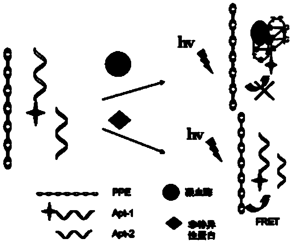
[0025] 1) Mix dye-labeled aptamer fragment 1, aptamer fragment 2 and water-soluble conjugated polymer at a molar ratio of 1:1:5 to form an aptamer / polymer complex, and then add it to the buffer solution In this method, the fluorescence detection is carried out with 404 nm as the excitation wavelength, the fluorescence emission band of the complex is recorded, and the ratio I of the fluorescence intensity at 525 nm to 440 nm is calculated 525 / I 440 ; Aptamer fragment 1 and aptamer fragment 2 are specific single nucleotide sequences, the 5' end of aptamer fragment 1 is labeled with fluorescein isothiocyanate, and the labeling method is: 5'-6-FITC.

[0026] 2) Add aptamer fragment 1 and aptamer fragment 2 and the test sample containing thrombin into the binding solution and place it at 37°C for 40 minutes to form a thrombin / aptamer complex.

[0027] 3) Adding a water-soluble conjugated polymer to the above complex, thrombin induces and combines with aptamer fragment 1 and aptam...

Embodiment 2

[0029]1) Mix aptamer fragment 1, dye-labeled aptamer fragment 2 and water-soluble conjugated polymer at a molar ratio of 1:1:10 to form an aptamer / polymer complex, and then add it to the buffer solution In this method, the fluorescence detection is carried out with 404 nm as the excitation wavelength, the fluorescence emission band of the complex is recorded, and the ratio I of the fluorescence intensity at 525 nm to 440 nm is calculated 525 / I 440 ; The aptamer fragment 1 and the aptamer fragment 2 are specific single nucleotide sequences, and the 3' end of the aptamer fragment 2 is labeled with fluorescein, and the labeling method is: 3'-6-FAM labeling.

[0030] 2) Add aptamer fragment 1 and aptamer fragment 2 and the test sample containing thrombin into the binding solution and place it at 37°C for 40 minutes to form a thrombin / aptamer complex.

[0031] 3) Adding a water-soluble conjugated polymer to the above complex, thrombin induces and combines with aptamer fragment 1 ...

Embodiment 3

[0032] Example 3 Specific Analysis of Thrombin Detection

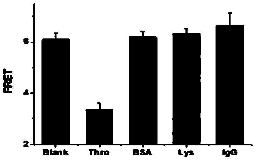
[0033] Add 2.5 μL of aptamer fragment 1 at a concentration of 10 μM and 2.5 μL of aptamer fragment 2 at a concentration of 10 μM to each centrifuge tube, then add 1 μL of thrombin (Thro) at a concentration of 10 μM, 6.8 μL Bovine serum albumin (BSA) at a concentration of 0.1 mg / ml, 2 μL of lysozyme (Lys) at a concentration of 5 μM, and 1.6 μL of immunoglobulin (IgG) at a concentration of 1 mg / ml, so that the final concentration of each protein was 20 nM, The final concentration of aptamer fragment 1 and aptamer fragment 2 is 50 nM; finally add 2.5 μL of a water-soluble conjugated polymer with a concentration of 50 μM, and perform fluorescence detection under the environment of pH=10.6, 0.1 M CBS buffer solution , see the result image 3 , it can be seen from the figure that the FRET of the blank and the addition of non-specific protein are similar and relatively high, and the FRET of the addition of thrombin is signif...


PUM


Abstract

The invention discloses a thrombin detection method based on a splitter adapter and a water-soluble conjugated polymer. According to the method, a super-molecular detection platform based on a water-soluble conjugated polymer/nucleic acid adapter is designed by virtue of the sensitivity of the water-soluble polymer on a comformational change of a deoxyribonucleic acid (DNA) chain and according to the characteristics that the nucleic acid adapter has specific binding ability and the like. Two sequences cannot form a G-four-chain structure when thrombin does not exist in a solution or other nonspecific proteins exist in the solution, the distance from a compound to the polymer is small, and thus effective energy transfer between the compound and the polymer can be realized; when the thrombin exists in the solution, the thrombin induces the two sequences to form the G-four-chain structure, and the distance between the compound and the polymer is increased, so that effective energy transfer cannot be carried out. The method disclosed by the invention is simple, convenient and fast, is high in sensitivity and selectivity, and has important significance in the aspect of thrombin detection.

Description

technical field [0001] The invention belongs to the field of biosensing and analysis, and particularly relates to a fluorescent detection method for detecting thrombin by using split aptamers and water-soluble conjugated polymers. Background technique [0002] Thrombin is a proteolytic enzyme formed from the precursor of thrombin (an essential component in plasma), which catalyzes the conversion of fibrinogen into fibrin to promote blood coagulation. The detection of thrombin is of great significance to the diagnosis of clinical diseases, the course of disease development, prognosis, and the monitoring and evaluation of curative effect. [0003] Since the thrombin produced in the early stage of blood coagulation is quickly neutralized by the anticoagulant substances in the body, it is very difficult to directly measure thrombin. Clinical thrombin detection mostly uses indirect detection methods, that is, to reflect the generation of thrombin by detecting specific coagulatio...

Claims


Application Information

Patent Timeline
Patent Type & Authority Applications(China)
IPC IPC(8): G01N21/64
Inventor 刘兴奋石琳黄艳琴黄维
Owner 深圳云安智慧医疗科技有限公司
Why Eureka
Try Eureka