Mouse uterus perfusion apparatus and method for establishing mouse endometritis model
An endometritis and perfusion device technology, applied in the medical field, can solve the problems of establishing a mouse endometritis model, difficulty in perfusion, perfusion fluid perfusion, etc., to reduce mechanical damage, shorten operation time, and reduce outflow Effect
- Summary
- Abstract
- Description
- Claims
- Application Information
AI Technical Summary
Problems solved by technology
Method used
Image
Examples
Embodiment 1
[0041] Example 1: Determination of the volume of perfusate in the mouse uterus
[0042] In order to determine the optimum volume of perfusate in the mouse uterus, this experiment perfused 10, 20, and 40 μl of PBS in the mouse uterus respectively, observed the performance of the mice after perfusion, and determined the volume of the perfusate in the mouse uterus. It was found that when the volume of perfusate in the uterus of each mouse was 40 μl, the perfusion was difficult and the vulva fluid flowed out after perfusion; if the perfusion was 10 μl, although the perfusion fluid could completely enter the uterus, the perfusion fluid could not be evenly distributed. Therefore, the volume of mouse uterine perfusate was finally determined to be 20 μl / mouse.
Embodiment 2
[0043] Embodiment 2: Comparison of anesthesia methods
[0044] In order to compare the effects of different general anesthesia methods on the establishment of this model, a comparative test of anesthesia was carried out with inhalation anesthetics such as ether and Sumianxin injection. Twelve mice were divided into ether anesthesia group and Sumianxin anesthesia group. Ether anesthetized group: place the mouse upside down in a beaker, and put a cotton ball soaked in ether into the beaker. Sumianxin anesthesia group: Sumianxin injection was injected into the rear thigh muscle of mice at a dose of 0.67ml / kg. After anesthesia, 20 μL lipopolysaccharide with a concentration of 2.5 mg / mL was perfused in the uterus, and the change of NO content in the uterus of mice was detected 24 hours after perfusion.
[0045] It takes 5-10 minutes for the mice to go from the injection of Sumianxin to general anesthesia, and the maintenance period can be maintained for 1-2 hours. However, the m...
Embodiment 3
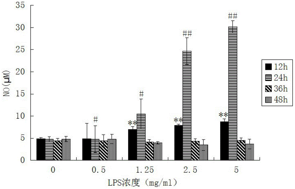
[0046] Example 3: Screening of LPS concentration and time in the mouse endometrium inflammation model
[0047] The key to the establishment of the mouse endometrial inflammation model is the concentration of the modeling drug and the time when the obvious inflammatory response appears. The appropriate concentration and time range can only be determined by designing concentration gradients and time gradients for screening.
[0048] 1.1 Test animals
[0049] 120 8-12-week-old clean-grade Kunming female mice (purchased from Qingdao Drug Control Institute), weighing 30-35 g. 12h of light alternates with 12h of darkness, the indoor temperature is 20-25°C, and feed and drinking water are given normally. After one week of adaptive feeding, it will be ready for use.
[0050] 1.2 Main reagents and kits
[0051] LPS (Sigma, O127: B8); Sumianxin Injection (Dunhua Shengda Animal Pharmaceutical Co., Ltd., Jilin Province, approval number: Veterinary Medicine (2010) 070031582); Myelopero...
PUM
| Property | Measurement | Unit |
|---|---|---|
| Length | aaaaa | aaaaa |
| Outer diameter | aaaaa | aaaaa |
| The inside diameter of | aaaaa | aaaaa |
Abstract
Description
Claims
Application Information
 - R&D
- Intellectual Property
- Life Sciences
- Materials
- Tech Scout
- Unparalleled Data Quality
- Higher Quality Content
- 60% Fewer Hallucinations
Browse by: Latest US Patents, China's latest patents, Technical Efficacy Thesaurus, Application Domain, Technology Topic, Popular Technical Reports.
© 2025 PatSnap. All rights reserved.Legal|Privacy policy|Modern Slavery Act Transparency Statement|Sitemap|About US| Contact US: help@patsnap.com


