3D printing percutaneous vertebral pedicle guide plate, preparation method of 3D printing percutaneous vertebral pedicle guide plate, and using method of 3D printing percutaneous vertebral pedicle guide plate

A 3D printing and pedicle technology, applied in the medical field, can solve the problem of rare mobile CT in the operating room, and achieve the effects of shortening the operation time, accurate screw placement, and simple structure

Active Publication Date: 2015-01-21
江苏舟可医疗器械科技有限公司
View PDF5 Cites 21 Cited by
  • Summary
  • Abstract
  • Description
  • Claims
  • Application Information

AI Technical Summary

Problems solved by technology

At present, there is a more advanced CT-guided pedicle screw placement technology, which can achie

Method used


Image

  • 3D printing percutaneous vertebral pedicle guide plate, preparation method of 3D printing percutaneous vertebral pedicle guide plate, and using method of 3D printing percutaneous vertebral pedicle guide plate
  • 3D printing percutaneous vertebral pedicle guide plate, preparation method of 3D printing percutaneous vertebral pedicle guide plate, and using method of 3D printing percutaneous vertebral pedicle guide plate
  • 3D printing percutaneous vertebral pedicle guide plate, preparation method of 3D printing percutaneous vertebral pedicle guide plate, and using method of 3D printing percutaneous vertebral pedicle guide plate

Examples


preparation example Construction

[0035] The preparation method of the 3D printed percutaneous pedicle guide plate of the present invention includes the steps of establishing a three-dimensional geometric model, determining the pedicle screw path, generating the guide plate, 3D printing and installing the sleeve, which are specifically:

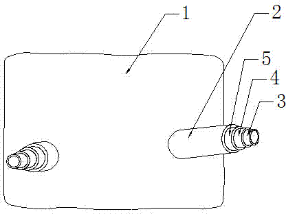
[0036] (1) The establishment of three-dimensional geometric model: Obtain the patient's imaging data from the workstation and import it into the medical modeling software in DICOM format. Through morphological processing, a visual bone model and skin model are generated, such as figure 2 with 3 As shown, and export and save in stl format respectively;

[0037] (2) Determination of the pedicle screw path: In the medical modeling software, extract the pedicle shape and fit into a cylindrical channel, which is the best screw path. Measure the length of the screw path in the software, and then The road is reversed and extended through the skin to form a body surface mark, and then exp...

specific example

[0049] A patient, male, 57 years old. He was admitted to the hospital for "repeated waist and hip pain for half a year". The physical examination showed that the lumbar spine had physiological arc, lumbar 4 and 5 spinous processes, interspinous tenderness, mild percussive pain, both lower limbs straight leg elevation test 70° negative, double Muscle strength and sensation of lower limbs are normal. Examination of the front and lateral view of the lumbar spine showed that the lumbar 4 vertebral body had second degree spondylolisthesis, and the lumbar spine CT showed that the lumbar 4th vertebral body had second degree spondylolisthesis, the intervertebral disc herniation was not obvious, and the pedicle was intact. The admission diagnosis was: "Lumbar 4 spondylolisthesis". Percutaneous pedicle screw internal fixation under general anesthesia was performed, and 3D printed percutaneous pedicle guide plates were used during the operation. Specific steps are as follows:

[0050] 1....


PUM


Abstract

The invention discloses a 3D printing percutaneous vertebral pedicle guide plate, a preparation method of the 3D printing percutaneous vertebral pedicle guide plate, and a using method of the 3D printing percutaneous vertebral pedicle guide plate. The guide plate comprises a wavy panel which can be attached to the spine of a human body, the panel is provided with two hollow input sleeves used for pedicle screws to penetrate through, and a first layer sleeve, a second layer sleeve and a third layer sleeve are sequentially arranged inside each inlet sleeve from inside to outside. The preparation method of the 3D printing percutaneous vertebral pedicle guide plate comprises the steps of the building of a three-dimensional geometrical model, the determination of pedicle screw channels, the generation of the guide plate, 3D printing and sleeve installation. The 3D printing percutaneous vertebral pedicle guide plate is simple in structure and convenient to prepare. In the preparation process of the guide plate, machining is carried out on the designed guide plate in advance according to a body surface mark so that locking can be achieved for body surface positioning, it is guaranteed that the inlet sleeves and the pedicle screw channels are coaxially arranged, displacement is prevented, and it is guaranteed that the direction of puncturing and the direction of screw placement are accurate; through the guide plate, the placement depth is accurate, fluoroscopy does not need to be carried out repeatedly, surgical accuracy is improved, and surgical time is shortened.

Description

Technical field [0001] The invention belongs to the field of medical technology, relates to a spinal surgery internal fixation operation device, in particular to a 3D printed percutaneous pedicle guide plate, and also relates to a preparation method and a use method of the guide plate. Background technique [0002] In the field of biomedicine, modern medicine tends to individualized treatment strategies. With the strong improvement of computer computing power, the extensive and in-depth combination of computer technology and medicine has formed a new interdisciplinary ---digital medicine, which has greatly promoted The development of modern clinical individualized diagnosis and treatment technology. At the same time, because 3D printing has the characteristics of individualization, it is consistent with the diagnosis and treatment concept of modern medicine. Therefore, it will be a match made in heaven to apply 3D printing technology to the field of biomedicine. At present, with...

Claims


Application Information

Patent Timeline
IPC IPC(8): A61B17/70
CPCA61B17/7083
Inventor 顾铁保王彭张泽锋
Owner 江苏舟可医疗器械科技有限公司
Why Eureka
Try Eureka