Method for determining activity of trichophyton rubrum keratinase

A keratinase activity, technology of Trichophyton rubrum, applied in the directions of microorganism-based methods, microorganism determination/inspection, biochemical equipment and methods, etc. Determining accurate effects

Active Publication Date: 2015-02-18
CHUGOKU IGAKU KAGAKUIN HIFUBIYOU KENKYUSHO
View PDF0 Cites 3 Cited by
  • Summary
  • Abstract
  • Description
  • Claims
  • Application Information

AI Technical Summary

Problems solved by technology

Clinically, the above-mentioned inducers do not exist when Trichophyton rubrum infects the human body, and there may be serious deviatio

Method used


Image

  • Method for determining activity of trichophyton rubrum keratinase
  • Method for determining activity of trichophyton rubrum keratinase

Examples


Embodiment 1

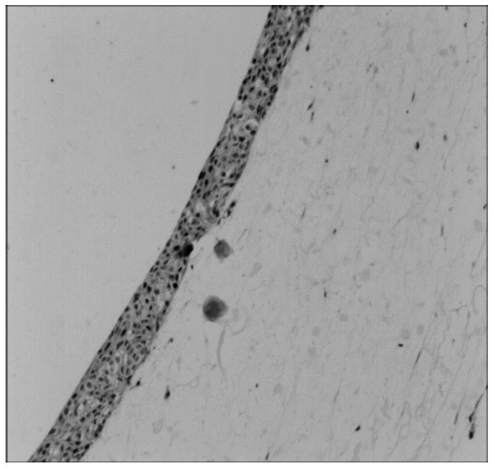
[0022] Under aseptic conditions, the 10 6 Personal skin fibroblasts were resuspended in 2ml of collagen solution, added dropwise to a 6-well cell culture plate, and placed at 37°C, 5% CO 2 cell culture incubator. After 3 days, it was observed under the microscope that the cells were fully expanded, and 10 6 The individual keratinocyte suspension was transferred to the surface of the collagen gel, and then placed in the cell incubator for culture. After 5 days, it was observed under the microscope that the human keratinocytes on the surface of the gel were completely covered, and the air-liquid surface culture was carried out. After that, HE staining of frozen sections was carried out every day, and the culture was stopped when the stratum corneum was differentiated to 5-8 layers after 10 days. Get the artificial skin containing epidermis and dermis structure (see figure 1 ).

Embodiment 2

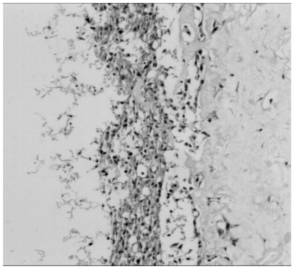
[0024] Resuspend the cultured Trichophyton rubrum in normal saline, adjust to 1 McFarland concentration (10 6 spores / ml), drop 5μl to the artificial skin containing epidermis and dermis structure prepared in step ⑴, and continue to culture for 48h. HE staining verified that Trichophyton rubrum successfully infected the artificial skin (see figure 2 ).

Embodiment 3

[0026] Collection step (2) About 4ml of the culture solution of the artificial skin infected with Trichophyton rubrum, 4000 rpm for 15 minutes, suck out 3ml of the supernatant, transfer it into 2ml of buffer solution containing 5mg of keratin-azure, put it in 200°C at 30°C Rpm shaker incubation. Replace the supernatant with 3 ml sterile water as a negative control. After 72 hours, place the reaction supernatant on an ice plate, absorb 1ml of the supernatant, and centrifuge at 12,000 rpm at 4°C for 5 minutes. Pipette the supernatant into a 96-well plate, 200 μl per well, repeat 3 wells, measure the OD value at A595 nm with a UV spectrophotometer, and use the blank solution containing the same amount of keratin-azure in sterile water as a negative control. Every 0.01 change in A595 nm compared with the negative control is equivalent to 1 U of keratinase activity, resulting in keratinase activity of 9.85±0.13 U.


PUM


Abstract

The invention relates to a method for determining the activity of a trichophyton rubrum keratinase. The method comprises the following steps: (1), constructing an artificial skin containing epidermis and dermis structures: resuspending human skin fibroblast cells in a collagen liquid, dropwise adding into a cell culture well plate and culturing; after the cells are completely expanded, dropwise adding a human keratinocyte suspension to the surface of collagen gel and continuously culturing; carrying out air-liquid interface culture when the human keratinocytes completely cover the surface of the collagen gel; and stopping culturing until a cuticular layer is differentiated into 5-8 layers; (2) carrying out artificial infection on trichophyton rubrum; and (3) determining the activity of the keratinase. The artificial skin, which is constructed by the method disclosed by the invention, becomes a natural inducer for production of the keratinase and has the following advantages that firstly, the artificial skin contains the dermal layer, the epidermal layer and 5-8 cuticular layers and the structure and function are closer to those of the human skin; secondly, keratin secreted by the keratinocyte in the epidermal layer is used as a natural substrate to induce the production of the keratinase; and, thirdly, the keratinase produced by inducing the natural substrate keratin, is accurate, real and stable in the determination of the activity.

Description

technical field [0001] The invention relates to a method for measuring the activity of Trichophyton rubrum keratinase, which belongs to the technical field of fungal microorganisms. Background technique [0002] Dermatophytes refer to fungi that can invade the stratum corneum and horny tissue, causing skin, hair and nail infections in humans and animals. It has been found that there are more than 40 kinds of dermatophytes, of which more than 30 are pathogenic. The most common dermatophytes that cause infection are Trichophyton rubrum, Trichophyton mentagrophytes, Microsporum canis, Microsporum gypsum, and Epidermophyton flocculus. Trichophyton rubrum is the most common pathogen causing dermatophytosis, and according to statistics, it can cause 90% of chronic refractory infections of dermatophytosis. [0003] Like other infectious diseases, the pathogenesis of dermatophytosis is the result of the interaction of fungal virulence factors with host barrier structures and the i...

Claims


Application Information

Patent Timeline
IPC IPC(8): C12Q1/37C12Q1/02C12R1/645
Inventor 刘维达马鹏程曹玉萍沈永年
Owner CHUGOKU IGAKU KAGAKUIN HIFUBIYOU KENKYUSHO
Why Eureka
Try Eureka