Fluorescent nano-particle based cell membrane imaging reagent and preparation method thereof

A fluorescent nanoparticle and fluorescence imaging technology, applied in the biological field, can solve the problems of fluorescent nanoparticle cell endocytosis, loss of cell membrane labeling specificity, etc., achieving good biocompatibility, excellent imaging effect, and saving staining time.

Active Publication Date: 2015-03-25
SOUTHEAST UNIV
View PDF2 Cites 7 Cited by
  • Summary
  • Abstract
  • Description
  • Claims
  • Application Information

AI Technical Summary

Problems solved by technology

However, most of the current fluorescent nanoparticles are used for the overall labeling of cells and the tracking of specific organelles, but not for the specific imaging of cell membranes. The main reason is that fluorescent nanoparticles themselves are easily endocytized by cells, thus losing cell membrane labels. specificity

Method used


Image

  • Fluorescent nano-particle based cell membrane imaging reagent and preparation method thereof
  • Fluorescent nano-particle based cell membrane imaging reagent and preparation method thereof
  • Fluorescent nano-particle based cell membrane imaging reagent and preparation method thereof

Examples


Embodiment 1

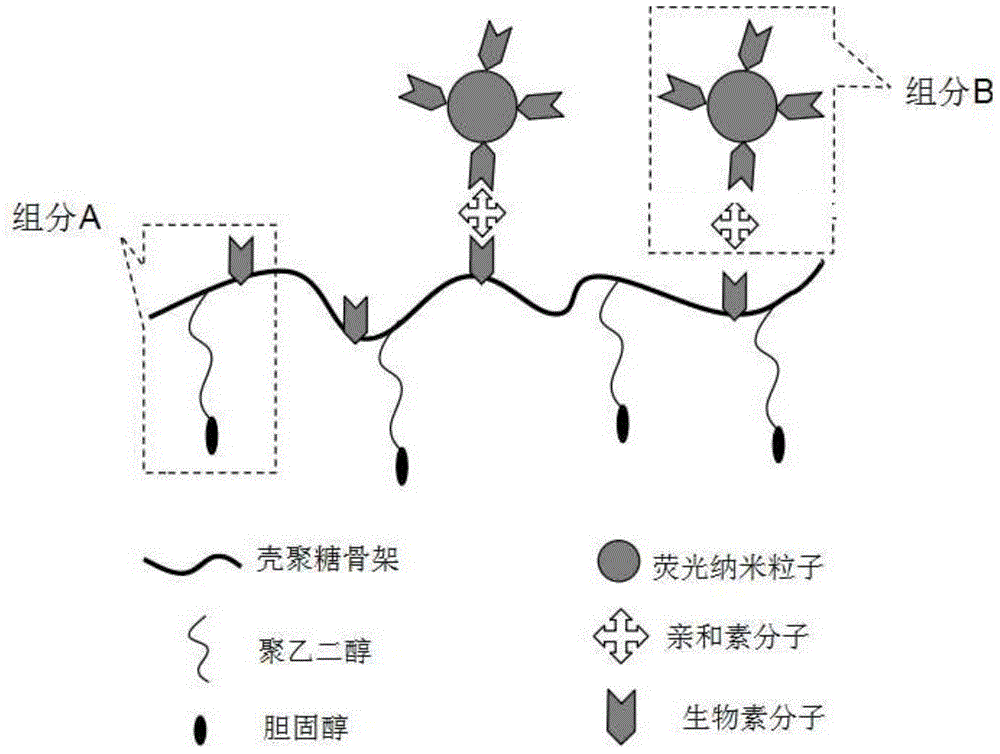
[0024] Cell membrane fluorescence imaging reagent based on fluorescent nanoparticles, composed of component A and component B. Component A: chitosan-10% cholesterol-10% biotin, component B: avidin-QDs (CdSeZnS).

[0025] Specifically, for the molecular structure of the fluorescent nanoparticle-based cell membrane fluorescence imaging reagent, see figure 1 , consisting of component A and component B. Among them, component A is based on glycol chitosan macromolecule (glycochitosan) as the backbone, and the side chain contains 10% polyethylene glycol 2000-cholesterol (PEG2000-cholesterol) hydrophobic units and 10% biotin molecules (biotin ); Ethylene glycol chitosan polymer has good biocompatibility and water solubility, the degree of polymerization is about 400, and the molecular weight is above 10,000. Experiments have proved that chitosan with a molecular weight of 10,000-100,000 can be well realized The present invention: the cholesterol hydrophobic anchoring unit is connec...

Embodiment 2

[0032] Cell membrane fluorescence imaging reagent based on fluorescent nanoparticles, composed of component A and component B. Component A: chitosan-10% cholesterol-10% biotin, component B1: avidin-biotin-CuS-FITC.

[0033] Specifically, for the molecular structure of the fluorescent nanoparticle-based cell membrane fluorescence imaging reagent, see figure 2 , consisting of component A and component B. Among them, component A is based on glycol chitosan macromolecule (glycochitosan) as the backbone, and the side chain contains 10% polyethylene glycol 2000-cholesterol (PEG2000-cholesterol) hydrophobic units and 10% biotin molecules (biotin ); ethylene glycol chitosan macromolecule has good biocompatibility and water solubility, the degree of polymerization is about 400, and the molecular weight is above 10,000; the hydrophobic side chain of cholesterol is connected to chitosan through PEG2000 with a molecular weight above 500 Amino groups, namely NHS-PEG2000-cholesterol, inc...

Embodiment 3

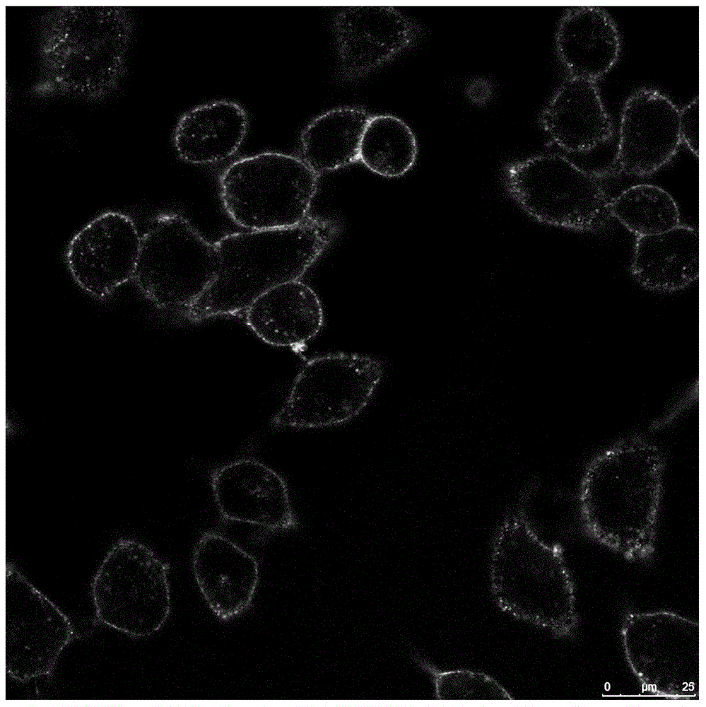
[0036] The implementation method of this embodiment is similar to the method in Example 2, except that component B is a silica nanoparticle doped with a rhodamine dye of grafted avidin, and the imaging effect is as follows Image 6 shown.


PUM

PropertyMeasurementUnit
degree of polymerizationaaaaaaaaaa

Abstract

The invention discloses a fluorescent nano-particle based cell membrane imaging reagent which comprises a component A and a component B, wherein the component A adopts an amino-contained polymer whose side chain contains a biotin molecule unit and a hydrophobic anchoring unit; and the component B adopts avidin module grafted fluorescent nano-particles. The reagent is based on fluorescent nano-particles and a multi-site anchoring technology and has good biocompatibility and good imaging effect, and the fluorescent particles are on a cell membrane, are not endocytosed, are not prone to quenching after long-time laser radiation and can be used for long-time tracing labeling of the cell membrane.

Description

technical field [0001] The invention belongs to the field of biotechnology, in particular to a cell membrane fluorescent imaging reagent based on fluorescent nanoparticles. Background technique [0002] The cell membrane not only serves as the boundary of the cell structurally, providing a relatively stable internal environment for the cell, but also plays a vital role in the process of material transportation, energy conversion and information transmission between the cell and the external environment. As a powerful research method, cell membrane imaging technology uses fluorescent dyes to label and trace the cell membrane, revealing many important biological processes including pinocytosis, exocytosis, cell division and apoptosis. [0003] However, some traditional lipophilic dyes not only endocytose quickly, but the imaging time is short; and these small organic molecule dyes are easy to be quenched (such as FITC and other organic fluorescent molecules will disappear afte...

Claims


Application Information

Patent Timeline
Patent Type & Authority Applications(China)
IPC IPC(8): G01N21/64
Inventor 吴富根王宏银贾浩然杨婧婧华先武
Owner SOUTHEAST UNIV
Why Eureka
Try Eureka