
Fluorescence biosensor for vascular endothelial growth factor detection in breast cancer

A biosensor and vascular endothelial technology, applied in the field of sensors, can solve the problems of high cost and cumbersome operation, and achieve the effect of strong ability, high sensitivity and huge application prospects

Inactive Publication Date: 2015-04-22
FUJIAN MEDICAL UNIV
View PDF1 Cites 8 Cited by
  • Summary
  • Abstract
  • Description
  • Claims
  • Application Information

AI Technical Summary

Problems solved by technology

The commonly used detection method is enzyme-linked immunoassay, but this method has the disadvantages of high cost and cumbersome operation, so it is necessary to find a more accurate, sensitive, economical, simple and non-invasive detection technology to detect this tumor Mark

Method used


Image

  • Fluorescence biosensor for vascular endothelial growth factor detection in breast cancer
  • Fluorescence biosensor for vascular endothelial growth factor detection in breast cancer
  • Fluorescence biosensor for vascular endothelial growth factor detection in breast cancer

Examples


Embodiment 1

[0023] The preparation method of fluorescent biosensor is as follows:

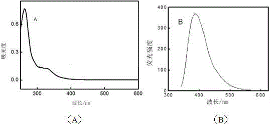
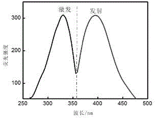
[0024] 1) Preparation of carbonized nitrogen nanomaterials: 2 mL of formamide was placed in a microwave tube, reacted at 170 °C for 30 min, washed 3 times with double distilled water, dried in vacuum for 8 h, dissolved in deionized water, and ultrasonically stripped for 24 h; The obtained liquid was centrifuged at 15000 r / min for 1 h, and the supernatant was obtained to obtain the suspension of carbonized nitrogen nanosheets; its spectrum was as follows: figure 1 and figure 2 shown;

[0025] 2) Preparation of 3-morpholine propanesulfonic acid (MOPS) buffer solution: weigh 0.5232 g of MOPS and 2.1248 g of NaNO 3 , after dissolving with double distilled water, transfer to 250 mL capacity, and constant volume, that is, 10 mmol / L MOPS solution; use 0.1 mol / L NaOH solution to adjust the pH to 7.0;

[0026] 3) Probe gene: 5'-CCCCCCTGTGGGGGTGGACGGGCCGGGTAGACCCCCC-3' (synthesized by Shanghai Sangon Bioenginee...

Embodiment 2

[0030] The fluorescence sensor of the present invention is used to detect VEGF, and the specific detection method is: adding a certain concentration of VEGF solution into the mixed system containing probe genes, silver ions and carbonized nitrogen nanosheets, and mixing and reacting. The above reaction solution was detected by fluorescence analysis.

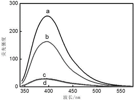
[0031] The present invention uses the fluorescence analysis method detection technology of prior art to observe the fluorescence signal change of carbonized nitrogen nanomaterial; Experimental result sees attached Figure 4, it can be seen from the figure that within a certain range, as the concentration of the target protein increases (from 10 pmol / L to 70 pmol / L), the probe gene of the hairpin structure combines with the protein to form a G-quadruplex structure and opens and releases The more silver ions are released, the stronger the fluorescence degree of quenched carbonized nitrogen, and the weaker the fluorescence signal; t...


PUM


Abstract

The invention belongs to the field of sensors, and particularly relates to a fluorescence biosensor for vascular endothelial growth factor detection in breast cancer. The fluorescence biosensor comprises nitrogen carbide nanometer materials, silver ions and a DNA probe. The gene of the DNA probe is 5'-CCCCCCTGTGGGGGTGGACGGGCCGGGTAGACCCCCC-3'. The silver ions and the DNA probe form a hairpin structure. Under the induction of a target molecule vascular endothelial growth factor, the NDA probe and target molecules are combined to form G-quadruplex structure, the silver ions are released, and fluorescence signals of the nitrogen carbide nanometer materials are quenched. When the fluorescence biosensor is used for early diagnosis of breast cancer, the detection limit of the fluorescence biosensor to the vascular endothelial growth factor is 3.5 pmol / L. When the fluorescence biosensor is used for clinical application, detection is more is more accurate, more sensitive, more economical, easier, more convenient and noninvasive, and the fluorescence biosensor is of great significance.

Description

technical field [0001] The invention belongs to the field of sensors, in particular to a fluorescent biosensor for detecting vascular endothelial growth factor in breast cancer. Background technique [0002] In recent years, with the development of new technologies such as molecular biology, the detection of tumor markers has attracted the attention of many scholars in the study of tumor diseases. Tumor markers refer to a class of substances synthesized and released by tumor cells or produced by host cells in response to tumors during tumorigenesis and proliferation, and often exist in the form of metabolites such as antigens, enzymes, and hormones. Studies have found that the occurrence and progression of breast cancer are closely related to vascular endothelial growth factor (VEGF), and detecting its level is of great significance for the monitoring, prognosis, and treatment of breast cancer. . Vascular endothelial growth factor (VEGF) is an important factor regulating a...

Claims


Application Information

Patent Timeline
Patent Type & Authority Applications(China)
IPC IPC(8): C12Q1/68G01N21/64
CPCC12Q1/6825C12Q1/6886C12Q2600/136C12Q2565/607C12Q2561/101
Inventor 李春艳陈敬华张静赵燕苹林佳兰建明吴芳韩志钟
Owner FUJIAN MEDICAL UNIV
Learn More