Method for detecting activity of uracil-DNA glycosylase (UDG) based on fluorescence amplification strategy of label-free non-enzyme DNA machines

A technology of glycosylase activity and uracil, which is applied in the field of protein qualitative and quantitative detection, can solve the problems of sensitive protease reaction conditions, poor test result reproducibility, interference signal amplification ability, etc., to achieve low impact, improve sensitivity, low The effect of background signals

Inactive Publication Date: 2015-05-20
SHANDONG UNIV
View PDF2 Cites 29 Cited by
  • Summary
  • Abstract
  • Description
  • Claims
  • Application Information

AI Technical Summary

Problems solved by technology

In addition, proteases are sensitive to reaction conditions and are prone to inactivation...

Method used


Image

  • Method for detecting activity of uracil-DNA glycosylase (UDG) based on fluorescence amplification strategy of label-free non-enzyme DNA machines
  • Method for detecting activity of uracil-DNA glycosylase (UDG) based on fluorescence amplification strategy of label-free non-enzyme DNA machines
  • Method for detecting activity of uracil-DNA glycosylase (UDG) based on fluorescence amplification strategy of label-free non-enzyme DNA machines

Examples


Embodiment 1

[0043] Example 1: Pure UDG activity detection

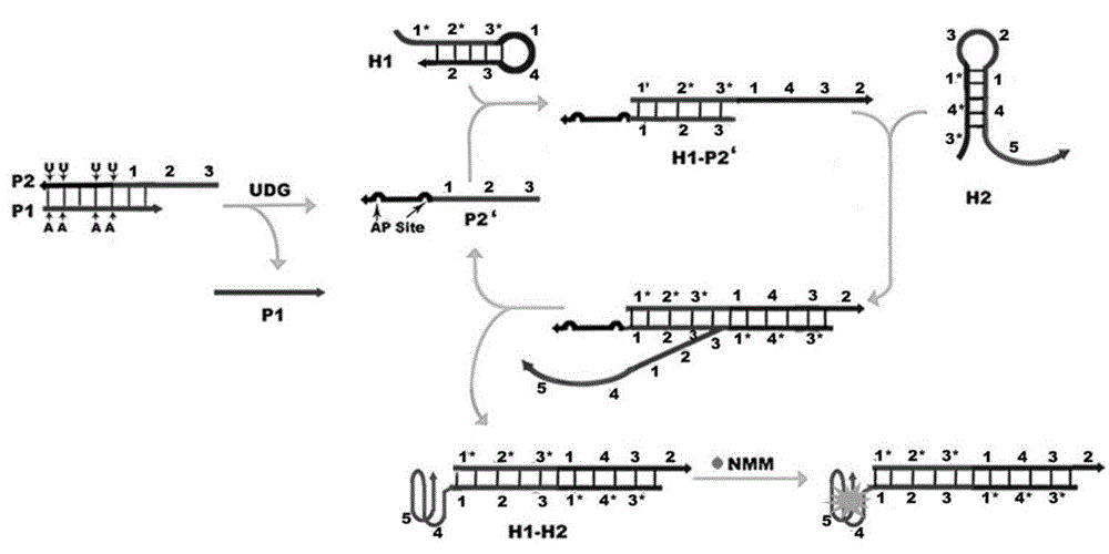
[0044] First design a double-stranded DNA (dsDNA) probe P1-P2 containing a uracil base and a priming sequence, wherein the P1 strand is an inhibitory strand, and its nucleotide sequence is as shown in SEQ ID NO.1 in the sequence listing; the P2 strand It is a chimeric conjugated strand containing a uracil DNA sequence and a priming sequence, and its nucleotide sequence is shown in SEQ ID NO.2 in the sequence table; the P1 strand and the P2 strand are partially complementary to form a double-stranded DNA (dsDNA) probe P1- P2;

[0045] P1: 5'-GAAATTCTTAAGTAGTCAG-3';

[0046] P2:5'-CGACATCTAACCTAGCTGACTACUUAAGAAUUTC-3'; (wherein, the part in italics is the triggering sequence)

[0047] According to the priming sequence of the P2 chain, two partially complementary hairpin probes H1 and H2 were designed for the construction of a label-free and enzyme-free DNA machine, and the G-quadruplex (G4) sequence was grafted on the end of the ...

Embodiment 2

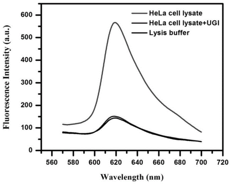
[0052] Example 2: Detection of UDG activity in cervical cancer (Hela) cell lysate

[0053] Adopt detection method of the present invention to analyze and detect the UDG activity in Hela cell lysate, concrete steps are as follows:

[0054] Hela cell samples were pelleted by centrifugation (5 min, 3000 rpm, 4°C) and redispersed in lysis buffer (10 mM Tris-Hcl, pH 7.0) by sonication on ice. The mixed solution was then centrifuged at 12,000 rpm for 30 min at 4°C to remove insoluble substances. The supernatant was collected and filtered through a 0.45 μm filter membrane to prepare a crude Hela cell lysate.

[0055] Crude Hela cell lysates can be directly assayed for UDG activity without further processing. The detection method is the same as the pure UDG activity detection method in Example 1.

[0056] In order to verify the applicability of the detection method of the present invention in the detection of UDG activity, we also used the method of the present invention to analyze...

Embodiment 3

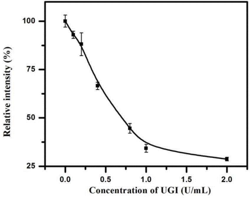
[0057] Example 3: Detection of activity inhibition of UDG

[0058] In order to detect the activity inhibition of UDG, the prepared dsDNA probes P1-P2 were mixed with 1 U / mL UDG and different concentrations of UGI, and incubated at 37°C for a certain period of time. Hairpins H1 and H2 were then added to the mixture to give a system with a final volume of 100 μL. At 37°C, the DNA machine performs amplification reactions for a certain period of time. Finally, NMM was added to the product and incubated at 37°C for 30min. All experiments were repeated three times.

[0059] UGI was chosen as a model inhibitor. UGI can form a tight and physiologically irreversible complex with UDG at a molar ratio of 1:1, such as image 3 As shown, when UGI exists, the relative fluorescence intensity of the system decreases with the increase of UGI concentration. The results demonstrate that this strategy can be used to detect the activity inhibition of UDG.


PUM


Abstract

The invention discloses a method for detecting the activity of uracil-DNA glycosylase (UDG) based on a fluorescence amplification strategy of label-free non-enzyme DNA machines, and the method comprises the following steps: (1) the preparation of a probe for the recognition and signal transduction of the UDG: firstly, designing an uracil base and initiation sequence containing double-stranded DNA probe P1-P2, wherein the P1 chain is an inhibition chain and the nucleotide sequence thereof is show in SEQ ID NO.1 in a sequence table; the P2 chain is a uracil-DNA sequence and initiation sequence containing chimeric conjugated chain and the nucleotide sequence thereof is show in SEQ ID NO.2 in the sequence table; and the P1 chain and the P2 chain are partially complemented so as to form the double-stranded DNA probe P1-P2; (2) the construction of a label-free non-enzyme DNA machine: according to an initiation sequence of the P2 chain, designing hairpin probes H1 and H2 which are partially complemented and used for constructing the label-free non-enzyme DNA machine, and grafting a G-quadruplet sequence to the tail end of the hairpin probe H2; and (3) the activity detection of UDG. The method disclosed by the invention successfully realizes background diminishing and signal amplification, and the LOD (limit of detection) is 0.00044 U/mL.

Description

technical field [0001] The invention relates to a method for detecting uracil-DNA glycosylase activity based on a label-free and enzyme-free DNA machine fluorescence amplification strategy, which belongs to the field of protein qualitative and quantitative detection. Background technique [0002] Uracil-DNA glycosylase (UDG) is a DNA repair enzyme that plays a key role in maintaining the integrity of the cellular genome. It removes uracil lesions from DNA by catalyzing the hydrolysis of the N-glycosidic bond connecting uracil to deoxyribose. Abnormal UDG activity will suppress the cellular response to uracil damage and is directly related to various diseases, including human immunodeficiency, lymphoma and Bloom syndrome. The findings suggest that UDG activity has become a promising biomarker for these diseases, and detection of UDG activity is a potential candidate tool in UDG functional studies and clinical diagnosis. [0003] Common methods for detecting UDG activity are...

Claims


Application Information

Patent Timeline
IPC IPC(8): C12Q1/68C12Q1/34
CPCC12Q1/682C12Q1/34
Inventor 姜玮王磊吴玉姝
Owner SHANDONG UNIV
Why Eureka
Try Eureka