Method for applying calixarene fluorescent probes to fluorescent imaging of Zn<2+> and F<->

A fluorescent probe and calixarene technology, applied in the field of analytical chemistry, achieves the effects of simple imaging method, good membrane penetration, and high luminescence yield

Active Publication Date: 2015-08-05
中知在线股份有限公司
View PDF4 Cites 8 Cited by
  • Summary
  • Abstract
  • Description
  • Claims
  • Application Information

AI Technical Summary

Problems solved by technology

In addition, among the fluorescent probes of thia and oxacalixarene, there are few reports that can be used for fluorescent staining of active cells, and can simultaneously realize the detection of trace Zn in cells. 2+ and F - The method of fluorescence imaging has not been reported
T...

Method used


Image

  • Method for applying calixarene fluorescent probes to fluorescent imaging of Zn&lt;2+&gt; and F&lt;-&gt;
  • Method for applying calixarene fluorescent probes to fluorescent imaging of Zn&lt;2+&gt; and F&lt;-&gt;
  • Method for applying calixarene fluorescent probes to fluorescent imaging of Zn&lt;2+&gt; and F&lt;-&gt;

Examples


Embodiment 1

[0040] Embodiment one: the preparation method of each solution, reagent among the present invention

[0041] (1) Preparation method of probe 1 and probe 2 solutions: weigh 13 mg of probe 1 and or probe 2, and use THF / H 2 O (THF accounted for 10% by volume) was dissolved and prepared into a 100mL solution with a concentration of 1.00×10 -4 mol L -1 ;

[0042] (2) Zn 2+ Standard solution: weigh 37mg of analytically pure Zn(ClO 4 ) 2 , dissolved in normal saline, and prepared into a 100mL solution with a concentration of 1.00×10 -3 mol L -1 ; Dilute step by step with normal saline to an appropriate concentration as needed;

[0043] (3)F - Standard solution: Weigh 26mg of analytically pure tetrabutylammonium fluoride, dissolve it in normal saline, and prepare a 100mL solution with a concentration of 1.00×10 -3 mol L -1 ; Dilute step by step with normal saline to an appropriate concentration as needed;

[0044] (4) 75% ethanol solution: Add 75mL of absolute ethanol to ...

Embodiment 2

[0051] Embodiment two: the culture of PC3 cell and HeLa cell

[0052] (1) Recovery cells

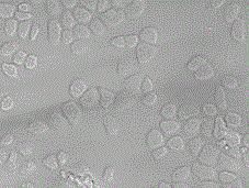
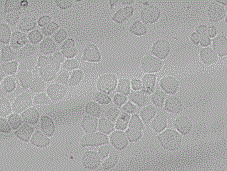
[0053] Take out the PC3 cells and HeLa cells from the -80°C refrigerator, place them in 37°C water and shake the cell cryopreservation tubes quickly, and thaw them completely within 1-2 minutes. 11ml of the culture solution prepared in Example 1 (PC3 cells plus improved RPMI-1640 culture solution, HeLa cells plus Gaotang DMEM culture solution) were mixed evenly, and the cell suspension was centrifuged at 1000r / min for 5min to remove For the supernatant, the cells precipitated at the bottom and the culture medium were lightly blown and mixed, and then transferred to the culture bottle, so that the volume of the culture solution in the culture bottle was within 5-7mL, and placed at 37°C, containing 5% CO 2 cultured in an incubator.

[0054] (2) Observation—passage—grafting

[0055] Change the culture medium once a day, and observe the growth of the cells under a microscope until the PC3...

Embodiment 3

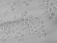
[0056] Example 3: Probe 1 to Zn in living PC3 cells 2+ , F - fluorescence microscopy

[0057] Group A: Add 10 μmol·L to 3 wells of the cell culture plate -1 Probe 1 solution (90% medium, 9% H 2 O, 1% THF, v / v) mixed solution; then add 20 μmol·L to the other 3 wells of the cell culture plate -1 Probe 1 solution (90% medium, 9% H 2 O, 1% THF, v / v) mixed solution at 37°C, containing 5% CO 2 Incubate in the incubator for 30 minutes, wash twice with fresh modified RPMI-1640 medium, place it under an IX-71 fluorescent inverted microscope for bright field and dark field imaging, and the cells show no fluorescence. (attached Picture 1-1 , 1-2 ). The PC3 cells in the above 6 wells incubated with probe 1 were immersed in a one-to-one correspondence with a concentration of 10 μmol L -1 Zn 2+ solution (90% medium, 10% H 2 O, v / v) in the mixed solution, 50 μmol L -1 Zn 2+ solution (90% medium, 10% H 2 O, v / v) in the mixed solution, 100 μmol L -1 Zn 2+ solution (90% medium, ...


PUM


Abstract

The invention discloses a method for applying calixarene fluorescent probes to fluorescent imaging of Zn<2+> and F<->. Two thiacalix[4]arene fluorescent probes selectively bonded with Zn<2+> and F<-> and having chemical names of 1,3-alternative-5,11,17,23-tetra-tert-butyl-25,27-di[(7-hydroxy-8-coumarinimino)ethoxy]-26,28-di(2-methoxyethoxy)thiacalix[4] and 1,3-alternative-5,11,17,23-tetra-tert-butyl-25,27-di[(7-hydroxy-8-coumarinimino)ethoxy]-26,28-thiacalix[4]crown-5 are respectively used as fluorescent imaging reagents for trace Zn<2+> and F<-> in active cancer cells in the human body; and the probes are compatible with the active cells and have permeability and nontoxicity. After the probes 1 and 2 are used to respectively dye Zn<2+> and F<-> in the cells, an inverted fluorescence microscope is used to detect shot clear blue fluorescent cell images.

Description

technical field [0001] The invention belongs to the field of analytical chemistry, specifically the application of calixarene fluorescent probes to Zn in living cells 2+ , F - imaging method. Background technique [0002] Live cell imaging technology is an important research method in the field of life sciences, which can explain a variety of life and physiological phenomena in living cells. With the development of life sciences, information research on active species in cells, cell signal transduction and cell apoptosis has become more and more in-depth. For the first time, researchers at the UC Davis Center for Biophotonics Science and Technology (CBST) have successfully imaged the motion of nanoscale compartments inside living tumor cells using fluorescent labeling. The research results hold the promise of future breakthroughs in understanding the molecular causes and biomechanics of cancer and a host of other diseases, as well as advancing neuroscience and stem cell r...

Claims


Application Information

Patent Timeline
IPC IPC(8): G01N21/64
Inventor 曾晞李丽曾莉朱勤牟兰
Owner 中知在线股份有限公司
Why Eureka
Try Eureka