Test strip for non-invasive diagnosis of laryngopharyngeal reflux disease and preparing and using method thereof

A test strip and throat technology, applied in the field of clinical diagnosis, can solve the problems of reducing detection sensitivity and accuracy, uneven thickness, poor sensitivity, etc., and achieve the effects of improving accuracy and convenience, reducing detection cost, and improving sensitivity

Active Publication Date: 2015-11-25
中国人民解放军总医院第六医学中心 +1
View PDF5 Cites 4 Cited by
  • Summary
  • Abstract
  • Description
  • Claims
  • Application Information

AI Technical Summary

Problems solved by technology

The scale diagnosis lacks specificity and accuracy; the 24-hour hypopharynx pH monitoring is an invasive test, which is not easily accepted by patients; the pepsin kit currently used to detect pepsin in saliva is used to diagnose laryngopharyngeal reflux, although it is a non-invasive test. However, it is a traditional enzyme-linked immunosorbent technique with poor sensitivity, and the number of patients with laryngopharyngeal reflux every day is limited, and the content of pepsin in saliva is relatively low. high rate
[0004] In addition, traditional enzyme detection reagent strips are generally a single detection unit, and for the detection of throat reflux disease, due to the uncertainty of gastric reflux, long-term interval detection is required, and the consumption of general reagent strips is relatively large.
In addition, the detection module of the general reagent strip is fixed on the bottom plate by colloid, and the thickness of the detection module is easy to be uneven when divided, which will affect the detection results to varying degrees, thereby reducing the sensitivity and accuracy of detection.

Method used


Image

  • Test strip for non-invasive diagnosis of laryngopharyngeal reflux disease and preparing and using method thereof
  • Test strip for non-invasive diagnosis of laryngopharyngeal reflux disease and preparing and using method thereof

Examples


preparation example Construction

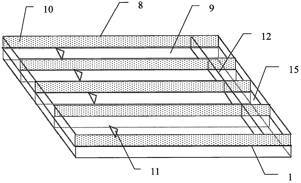
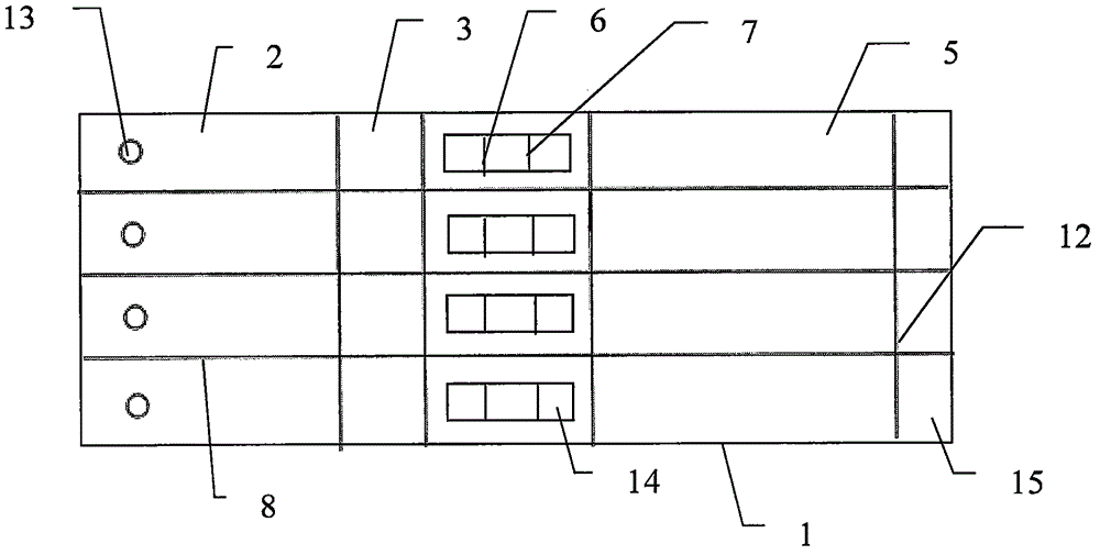
[0062] The preparation method of pepsin test strip for non-invasive diagnosis of throat reflux disease includes the following steps:

[0063] Step 1. Purify pepsin monoclonal antibody I and pepsin monoclonal antibody II;

[0064] Step 2. Prepare the binding pad 3. Covalently label the pepsin monoclonal antibody I obtained in step 1 on the gold nanoparticles, and then spray it on the glass fiber membrane, 35~38 under dark conditions. Dry at ℃ to obtain bonding pad 3;

[0065] Step 3. Prepare the detection pad. Use a phosphate buffer solution containing 4-8% sucrose to dilute the pepsin monoclonal antibody II and goat anti-mouse IgG antibody to a final concentration of 0.8-1.2 mg / ml, and then spray them in nitric acid at intervals. Dry the cellulose film to obtain the detection pad;

[0066] Step 4: Prepare the sample pad, soak the glass cellulose membrane with phosphate buffer solution for 20-40 minutes, and dry at 35-38°C to obtain the sample pad;

[0067] Step 5. Assemble. The detect...

Embodiment 1

[0080] Step 1. Purify pepsin monoclonal antibody I and pepsin monoclonal antibody II. Ion exchange chromatography and gel filtration techniques are used to separate and purify pepsin in human gastric juice; the purified pepsin is immunized against BALB / c mice, and monoclonal antibodies against human pepsin are prepared by hybridoma fusion technology; Chromatography and gel filtration technology are used to purify monoclonal antibodies, and enzyme-linked immunosorbent technology is used to screen highly specific diagnostic monoclonal antibodies.

[0081] Step two, prepare bonding pad 3. The gold nanoparticles are prepared by selecting and preparing gold nanoparticles with different diameters, and the prepared gold nanoparticles are characterized by ultraviolet-visible light analysis and transmission electron microscope analysis; firstly, the prepared gold nanoparticles are obtained by covalent coupling. The pepsin monoclonal antibody I was labeled on the gold nanoparticles. Then...

Embodiment 2

[0087] Step 1. Purify pepsin monoclonal antibody I and pepsin monoclonal antibody II. Ion exchange chromatography and gel filtration techniques are used to separate and purify pepsin in human gastric juice; the purified pepsin is immunized against BALB / c mice, and monoclonal antibodies against human pepsin are prepared by hybridoma fusion technology; Chromatography and gel filtration technology are used to purify monoclonal antibodies, and enzyme-linked immunosorbent technology is used to screen highly specific diagnostic monoclonal antibodies.

[0088] Step two, prepare bonding pad 3. The gold nanoparticles are prepared by selecting and preparing gold nanoparticles with different diameters, and the prepared gold nanoparticles are characterized by ultraviolet-visible light analysis and transmission electron microscope analysis; firstly, the prepared gold nanoparticles are obtained by covalent coupling. The pepsin monoclonal antibody I was labeled on the gold nanoparticles. Then...


PUM


Abstract

The invention discloses a test strip for non-invasive diagnosis of a laryngopharyngeal reflux disease and a preparing and using method thereof. The test strip comprises a base plate and a plurality of detection modules. The upper face and the lower face of the base plate are each provided with a plurality of parallel first partition plates, independent parallel grooves are formed, and the side walls of the grooves are provided with protruding thorns. Each detection module is formed by sequentially arranging and connecting a sample pad, a combined pad, a detection pad and water absorption paper in the length direction of the base plate, wherein the combined pad is coated with gold nanoparticles labeled with a pepsase monoclonal antibody I, and detection belts composed of pepsase monoclonal antibodies II and quality control belts composed of goat anti-mouse lgG antibodies are arranged on the detection pad at intervals. The detection modules are fixed in the different grooves respectively. Throat secretions or saliva is used as sample liquid to be detected, and the sample liquid to be detected is dropped to a sample adding hole; whether color development occurs on the quality control belts or not is continuously observed every two hours in 24 hours, and a patient does not have the laryngopharyngeal reflux disease if a color development reaction occurs.

Description

Technical field [0001] The present invention relates to the technical field of clinical diagnosis. More specifically, the present invention relates to a test strip for non-invasive diagnosis of throat reflux disease and a preparation and use method thereof. Background technique [0002] Throat reflux disease refers to the reflux of stomach contents above the upper esophageal sphincter, causing a series of symptoms and signs. The clinical manifestations are hoarseness or dysphonia, sore throat, foreign body sensation in the throat, continuous throat clearing, Chronic long-term cough, dyspnea, laryngospasm and other symptoms, as well as hyperplasia and hypertrophy of the mucous membrane in the commissural area of ​​the vocal cords, diffuse congestion and edema of the vocal cords, and severe throat signs such as granuloma, disappearance of the laryngeal compartment, and subglottic stenosis. [0003] Throat reflux disease is the source of some diseases in otolaryngology, head and neck ...

Claims


Application Information

Patent Timeline
IPC IPC(8): G01N33/577
CPCG01N33/577
Inventor 李进让吴立峰郭春雨
Owner 中国人民解放军总医院第六医学中心
Why Eureka
Try Eureka