Endoscopic oct imaging system using optical path difference external compensation common path interference probe

An imaging system and optical path difference technology, applied in the fields of endoscopy, application, medical science, etc., can solve the problems of difficult to enter the body, not traversal scanning imaging, difficult to practical needs, etc., to reduce the missed detection of lesions, resist the environment The effect of strong interference ability

Active Publication Date: 2017-11-24
INST OF OPTICS & ELECTRONICS - CHINESE ACAD OF SCI
View PDF7 Cites 0 Cited by
  • Summary
  • Abstract
  • Description
  • Claims
  • Application Information

AI Technical Summary

Problems solved by technology

The problems existing in these systems are as follows: 1) Only one imaging mode of spectral domain OCT is used, and there is no possibility of adopting frequency-sweeping OCT imaging mode, so different imaging modes cannot be used according to different application occasions to give full play to their respective advantages; 2) It is not traversing scanning imaging, and there is a possibility of missed detection of lesions; 3) The serious astigmatism problem that will be caused when the beam of incident sample passes through the cylindrical transparent sealing sleeve is not considered
[0005] In the frequency-sweeping OCT system proposed by Yang Yaliang et al. (Yang Yaliang et al., helical scanning co-path interference type endoscopic frequency-sweeping OCT real-time imaging method and system, invention patent: ZL2012100277848), the spectroscopic device and reference mirror are placed on the probe Internally, a common path interference endoscopic probe is formed, and a micro motor installed inside the probe is used to realize the helical scanning imaging at the top. The advantage of the top scanning is that it is stable, but the system still has the following problems: 1) Spectroscopic devices, reference mirrors, and the distance between the two to match the optical path of the sample beam, as well as the micro-motor, etc., make the unbendable part of the probe tip longer, which will make it difficult for the probe to pass through the oral cavity, the Y-shaped entrance of the electronic endoscope instrument channel, etc., so that it is not easy to into the body; 2) The micro-motor only performs helical scanning motion in the inner space of the tip of the probe, and the travel is very limited, which is difficult to meet the actual needs of lesion detection; 3) Only one imaging mode of frequency-sweeping OCT is used, and no spectral imaging mode is provided. 4) The astigmatism problem caused by the light beam passing through the cylindrical transparent sealing sleeve is not considered

Method used


Image

  • Endoscopic oct imaging system using optical path difference external compensation common path interference probe
  • Endoscopic oct imaging system using optical path difference external compensation common path interference probe
  • Endoscopic oct imaging system using optical path difference external compensation common path interference probe

Examples


Embodiment Construction

[0031] The present invention will be further described below in conjunction with the accompanying drawings and specific embodiments.

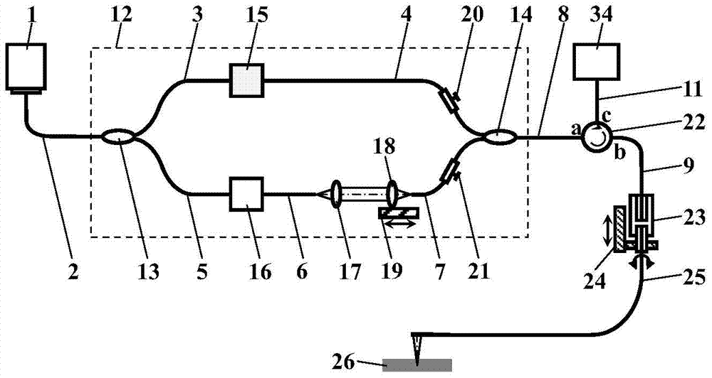
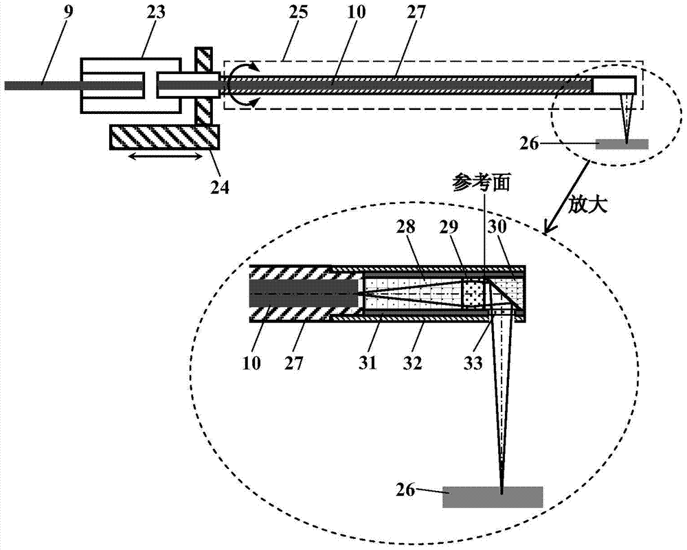
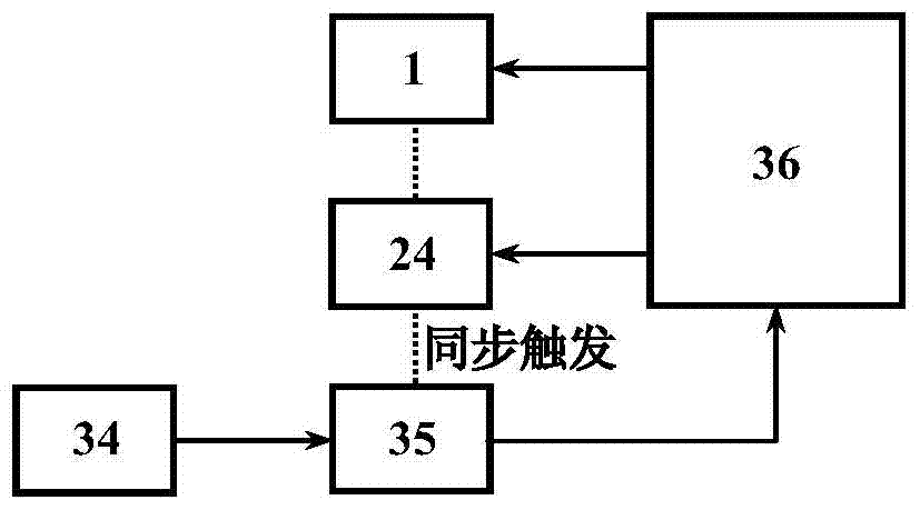
[0032] figure 1 It is a structural schematic diagram of an endoscopic OCT imaging system using an optical path difference externally compensated common path interference probe proposed by the present invention. Including: light source 1, first to tenth single-mode optical fiber 2-11, optical path difference external compensation interferometer 12, first and second fiber coupler 13-14, first and second acousto-optic frequency shifter 15- 16. First and second lenses 17-18, translation stage 19, first and second polarization controllers 20-21, optical circulator 22, optical fiber rotary connector 23, helical scanning mechanism 24, endoscopic probe 25, metal Coil 27, glass spacer rod 28, Green lens 29, 45° cylindrical reflector 30, transparent sealing sleeve 31, metal outer sheath 32, window 33, detector 34, data acquisition card 35, and computer ...


PUM


Abstract

The invention discloses an endoscopic OCT imaging system adopting an optical path difference external compensation common-path interference probe, comprising a light source, an optical path difference external compensation interferometer, first and second acousto-optic frequency shifters, an optical circulator, and an optical fiber Rotary connector, spiral scanning mechanism, endoscopic probe, glass spacer rod, Green lens, 45° cylindrical mirror, transparent sealing sleeve, metal outer sheath, window, detector, data acquisition card and computer, etc. Use the front surface of the Rigling lens of the endoscopic probe as the reference surface, and the optical path difference between it and the sample is compensated by the optical path difference external compensation interferometer; the spiral traversal scanning method is used to reduce the missed detection of lesions, and the flat window is used to To eliminate the astigmatism usually caused by the cylindrical transparent sealing sleeve, the acousto-optic frequency shifter combined with signal filtering and demodulation technology is used to eliminate the artifacts in the imaging results. The invention has the characteristics of strong anti-interference ability, plug-and-play probe, reduced missed detection of lesions, no false image interference, no astigmatism in incident sample beams, and the like.

Description

technical field [0001] The invention relates to medical endoscopic imaging technology and optical coherence tomography imaging technology, in particular to an endoscopic optical coherence tomography imaging system adopting optical path difference external compensation common path interference endoscopic probe and helical scanning imaging mode. Background technique [0002] Through the endoscope, doctors can directly observe the tissue morphology and lesions of the internal organs of the human body, and can also perform surgical treatment on the lesions. Traditional endoscopes can only observe the surface morphology of internal organs, and lesions often occur inside the organs first. Many imaging techniques with tomographic capabilities, such as: computer tomography, nuclear magnetic resonance, X-ray and ultrasound, are playing an important role in clinical diagnosis, but these techniques can only provide 0.1-1mm resolution, which is far from the detection The level of resol...

Claims


Application Information

Patent Timeline
Patent Type & Authority Patents(China)
IPC IPC(8): A61B5/00A61B1/07A61B1/00
Inventor 杨亚良张雨东
Owner INST OF OPTICS & ELECTRONICS - CHINESE ACAD OF SCI
Why Eureka
Try Eureka