A method for isolating and culturing skin keratinocytes
The technology of a keratinocyte and separation method is applied in the field of separation and acquisition of skin keratinocytes, which can solve the problems of rejection reaction and low efficiency, and achieve the effect of avoiding pollution and high vitality.
- Summary
- Abstract
- Description
- Claims
- Application Information
AI Technical Summary
Problems solved by technology
Method used
Image
Examples
Embodiment 1
[0061] Example 1: Successful isolation and expansion of human epidermal cells from adult normal chest skin
[0062] (1) Materials and methods
[0063] Sample: adult normal chest skin (including epidermis and dermis) discarded during hospital surgery, about 3cm 2 .
[0064] Cell culture medium: KGM-Gold.
[0065] (2) Cell isolation and culture
[0066] a) The parts where the skin is taken, the parts with hair should be removed first, and the cuticle should be removed in advance if the cuticle is too thick, and then sterilized with 75% alcohol cotton balls, and the tissue slices were collected.
[0067] b) Fresh skin samples are placed in HBSS / DPBS / DMEM, stored at 4°C and transported to the laboratory, and the area is measured.
[0068] c) Disinfect the sample in iodine tincture and 75% alcohol respectively, and then wash it with DPBS.
[0069] d) removing the yellow fat layer and most of the white dermis in the tissue sheet, retaining the epidermis and the 0.2-2 mm thick d...
Embodiment 2
[0078] Example 2: Successful isolation and expansion of human epidermal cells from adult normal chest skin
[0079] (1) Materials and methods
[0080] Sample: adult normal chest skin (including epidermis and dermis) discarded during hospital surgery, about 3cm 2 .
[0081] Cell culture medium: DKSFM.
[0082] (2) Cell isolation and culture
[0083] a) The parts where the skin is taken, the parts with hair should be removed first, and the cuticle should be removed in advance if the cuticle is too thick, and then sterilized with 75% alcohol cotton balls, and the tissue slices were collected.
[0084] b) Fresh skin samples are placed in HBSS / DPBS / DMEM, stored at 4°C and transported to the laboratory, and the area is measured.
[0085] c) Disinfect the sample in iodine tincture and 75% alcohol respectively, and then wash it with DPBS.
[0086] d) removing the yellow fat layer and most of the white dermis in the tissue sheet, retaining the epidermis and the 0.2-2 mm thick derm...
example 2
[0094] Example 2: Preparation of epidermal cells from healthy male foreskin
[0095] (1) Materials and methods
[0096] Sample: circumcised skin from a healthy male
[0097] (2) Cell separation and cultivation: the method is the same as in Example 1.
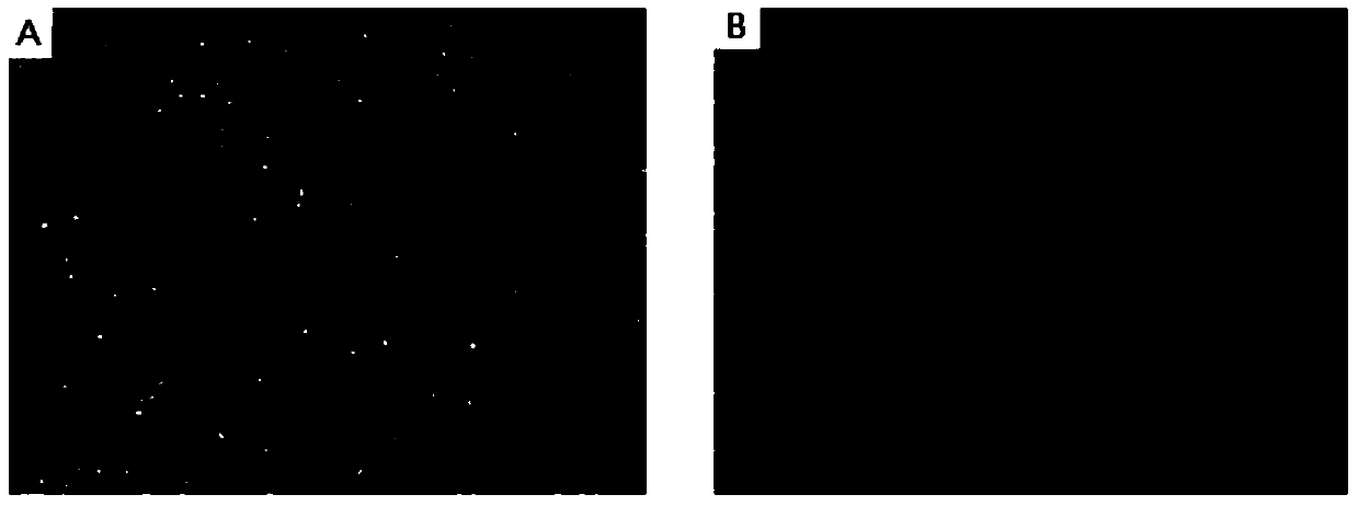
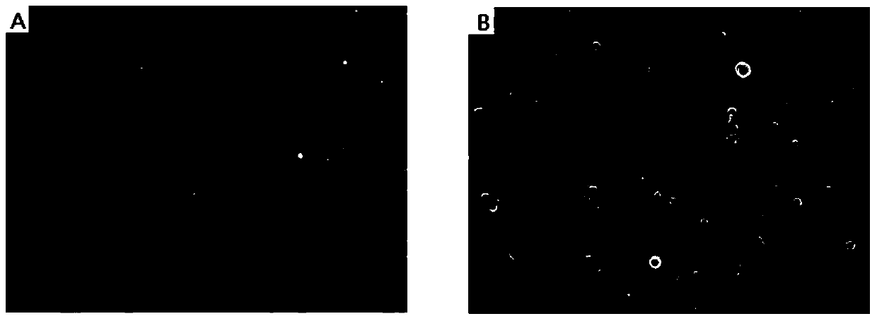
[0098] (3) Results: The isolated cells are easy to adhere to the wall, have more clones, and can quickly connect into sheets in the shape of "paving stones". Figure 4 shown. The cells are relatively pure, and can still maintain a good state and proliferation ability after passage and cryopreservation.
PUM
| Property | Measurement | Unit |
|---|---|---|
| thickness | aaaaa | aaaaa |
| thickness | aaaaa | aaaaa |
Abstract
Description
Claims
Application Information
 - R&D
- Intellectual Property
- Life Sciences
- Materials
- Tech Scout
- Unparalleled Data Quality
- Higher Quality Content
- 60% Fewer Hallucinations
Browse by: Latest US Patents, China's latest patents, Technical Efficacy Thesaurus, Application Domain, Technology Topic, Popular Technical Reports.
© 2025 PatSnap. All rights reserved.Legal|Privacy policy|Modern Slavery Act Transparency Statement|Sitemap|About US| Contact US: help@patsnap.com


