Preparation method of scanning electron microscope sample for polymer vesicae
A scanning electron microscope and polymer technology, which is applied in the detection field, can solve the problems of polymer vesicle morphology, size information distortion, and influence on observation, and achieve the effects of easy-to-obtain dyes, easy focus adjustment, and high image resolution
- Summary
- Abstract
- Description
- Claims
- Application Information
AI Technical Summary
Problems solved by technology
Method used
Image

Examples
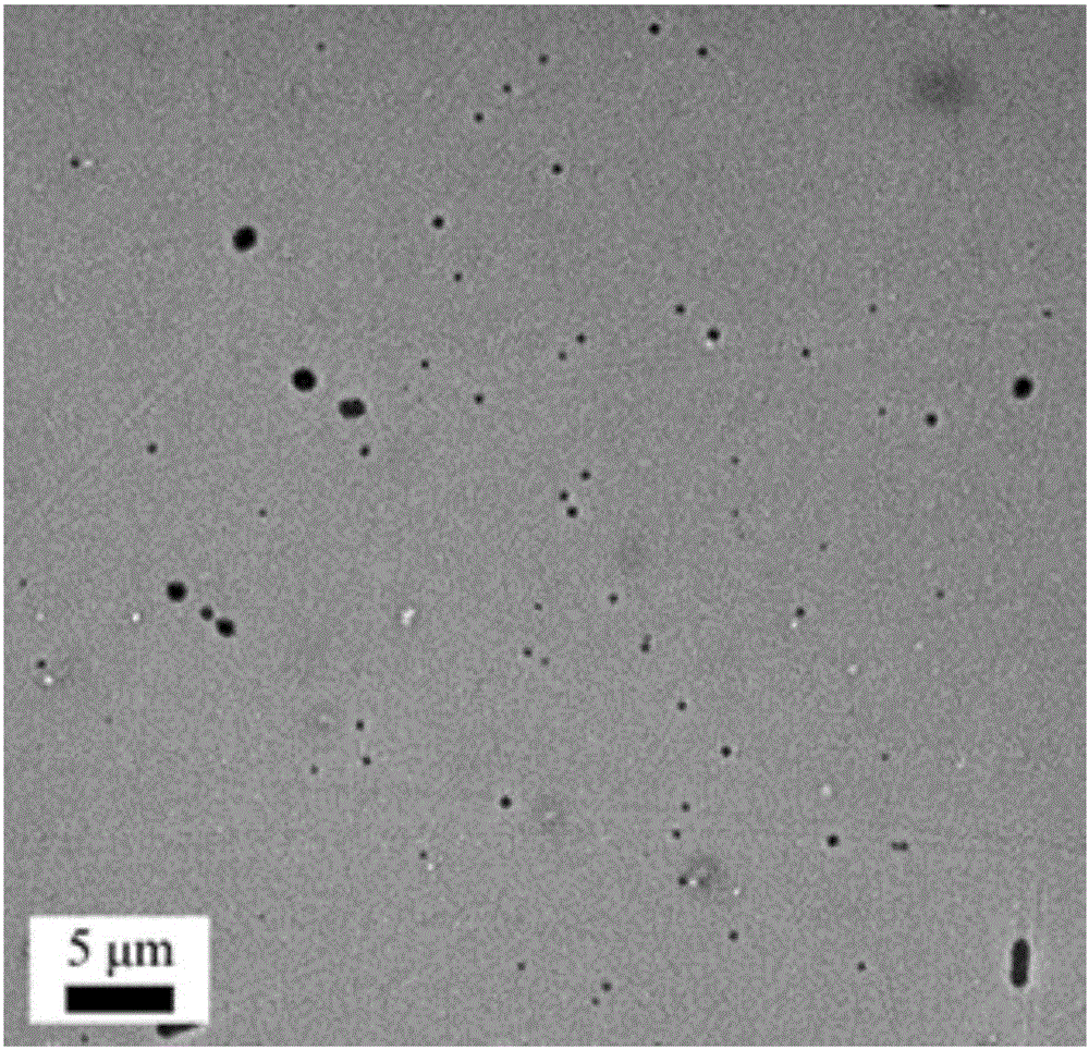
Embodiment 1
[0027] (1) Preparation of dye solution: Weigh 37.5mg of sodium phosphotungstate and add it to 5mL of water, and ultrasonicate for 10 minutes (ultrasonic power is 50W, ultrasonic frequency is 300kHz) to completely dissolve it to obtain phosphotungsten with a concentration of 0.75% w / w Sodium acid stain solution.
[0028] (2) Sample stage preparation: Paste conductive glue on the aluminum circular sample stage of the scanning electron microscope, and then attach a circular slide glass with a diameter of 12 mm.
[0029] (3) Staining and drying of samples: drop 50 μL PEG-PCL block copolymer nanoscale polymer vesicle solution on the glass slide, and then add 10 μL dye solution dropwise; use filter paper to lightly touch and absorb the excess mixed solution, The substrate is covered with a thin layer of mixed solution, and dried at room temperature to form a film to obtain a scanning electron microscope sample, at which point scanning electron microscopy can be photographed. The sc...
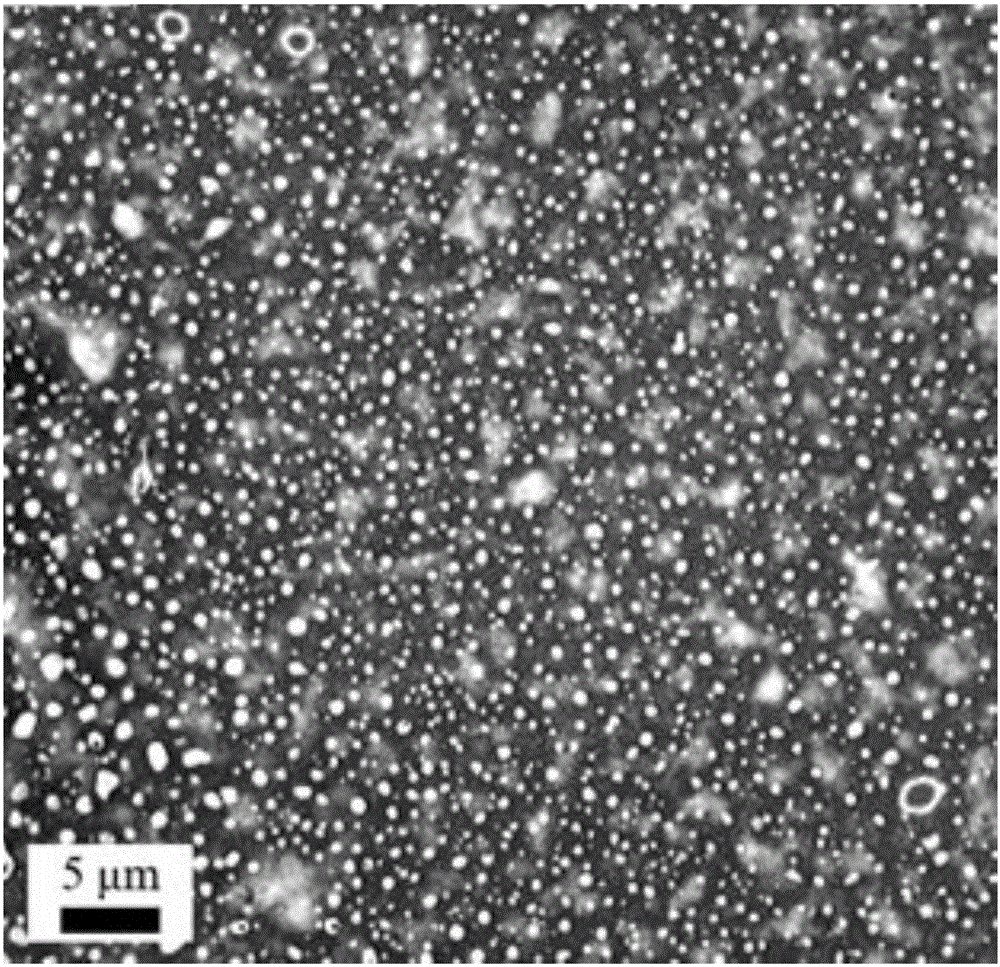
Embodiment 2
[0031] (1) Preparation of dye solution: Weigh 12.5mg of sodium phosphotungstate and add it to 5mL of water, and ultrasonicate for 5 minutes (ultrasonic power is 60W, ultrasonic frequency is 300kHz) to completely dissolve it to obtain phosphotungsten with a concentration of 0.25% w / w Sodium acid stain solution.
[0032] (2) Sample stage preparation: Paste conductive glue on the aluminum circular sample stage of the scanning electron microscope, and then attach a mica sheet with a diameter slightly smaller than the area of the sample stage.
[0033] (3) Staining and drying of samples: drop 50 μL PEG-PCL block copolymer nanoscale polymer vesicle solution on the glass slide, and then add 10 μL dye solution dropwise; use filter paper to lightly touch and absorb the excess mixed solution, The substrate is covered with a thin layer of mixed solution, and dried at room temperature to form a film to obtain a scanning electron microscope sample, at which point scanning electron micros...
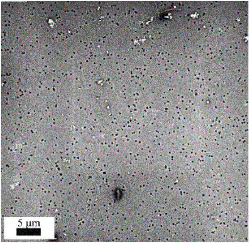
Embodiment 3
[0035] (1) Preparation of dye solution: Weigh 37.5mg of sodium phosphotungstate and add it to 5mL of water, stir evenly (stirring speed is 500r / min, stirring time is 30min), and obtain sodium phosphotungstate dyeing with a concentration of 0.75% w / w agent solution.
[0036] (2) Sample stage preparation: Paste conductive glue on the aluminum circular sample stage of the scanning electron microscope, and then attach a piece of aluminum foil with a diameter slightly smaller than the area of the sample stage.
[0037] (3) Sample staining and drying: Drop 50 μL of nanoscale polymer vesicle solution composed of PEG-PLA block copolymer on the glass slide, and then add 10 μL of dye solution dropwise; use filter paper to lightly touch and absorb the excess mixed solution, The substrate is covered with a thin layer of mixed solution, and dried at room temperature to form a film to obtain a scanning electron microscope sample, at which point scanning electron microscopy can be photogra...
PUM
 Abstract
Description
Claims
Application Information
 - R&D
- Intellectual Property
- Life Sciences
- Materials
- Tech Scout
- Unparalleled Data Quality
- Higher Quality Content
- 60% Fewer Hallucinations
Browse by: Latest US Patents, China's latest patents, Technical Efficacy Thesaurus, Application Domain, Technology Topic, Popular Technical Reports.
© 2025 PatSnap. All rights reserved.Legal|Privacy policy|Modern Slavery Act Transparency Statement|Sitemap|About US| Contact US: help@patsnap.com