Preparation and application method for liquid fluorescent microsphere with cell tracing function

A technology of fluorescent microspheres and cells, applied in chemical instruments and methods, preparations for in vivo experiments, luminescent materials, etc., can solve the problem of unfavorable dynamic observation of the same individual, and the inability to obtain cell location, viability, migration, and functional status in time and other problems, to achieve the effect of simple setting, easy practical operation and good light stability

Active Publication Date: 2016-07-20
HARBIN ENG UNIV
View PDF0 Cites 4 Cited by
  • Summary
  • Abstract
  • Description
  • Claims
  • Application Information

AI Technical Summary

Problems solved by technology

[0002] Although the traditional cell tracking method can obtain the information of the moment when the animal is put to death, it cannot obtain the location, viabili

Method used


Image

  • Preparation and application method for liquid fluorescent microsphere with cell tracing function
  • Preparation and application method for liquid fluorescent microsphere with cell tracing function

Examples


Embodiment Construction

[0027] The following examples describe the present invention in more detail.

[0028] (1) 1 μg nanocrystalline NaYF 4 :Yb 3+ ,Er 3+ Add it to 1mL organic solvent cyclohexane, put the mixed solution into an ultrasonic cleaner for ultrasonic treatment for one hour, so that the nanocrystals are uniformly dispersed in cyclohexane;

[0029] (2) Add 1 mL of organic solvent dioctyl ester to the solution after ultrasonication, and continue to put the mixed solution into an ultrasonic cleaner for one hour of ultrasonication. During this process, the weighing bottle containing the mixed solution should be opened to fully volatilize the cyclohexane. Obtain a dioctyl ester solution in which nanocrystals are uniformly dispersed;

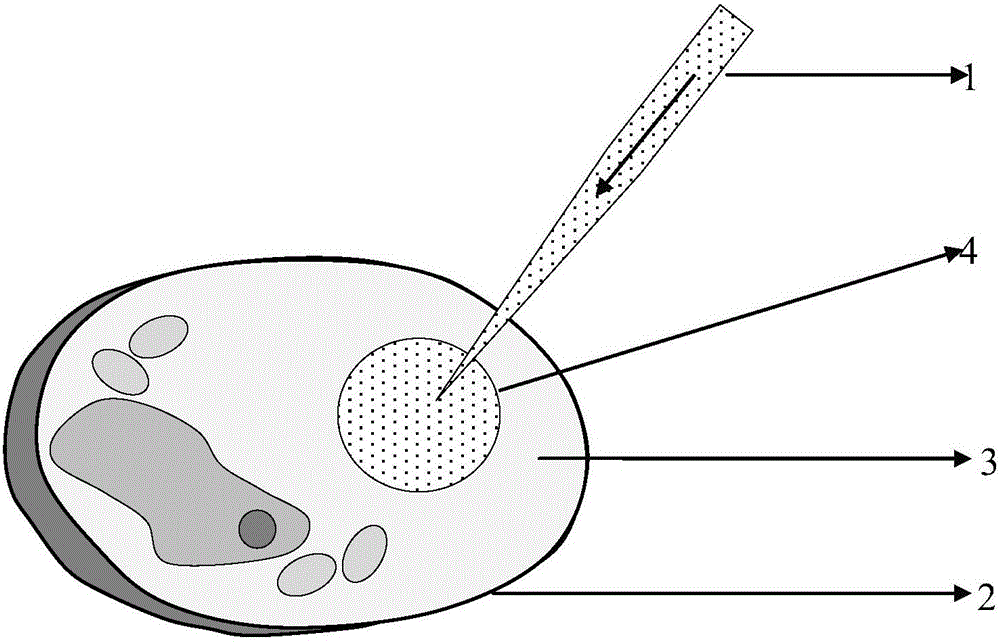
[0030] (3) A glass capillary injection tube with an outer diameter of 125 μm and an inner diameter of 100 μm is drawn into a tapered microneedle 1 with an inner diameter of 10 μm using a needle puller;

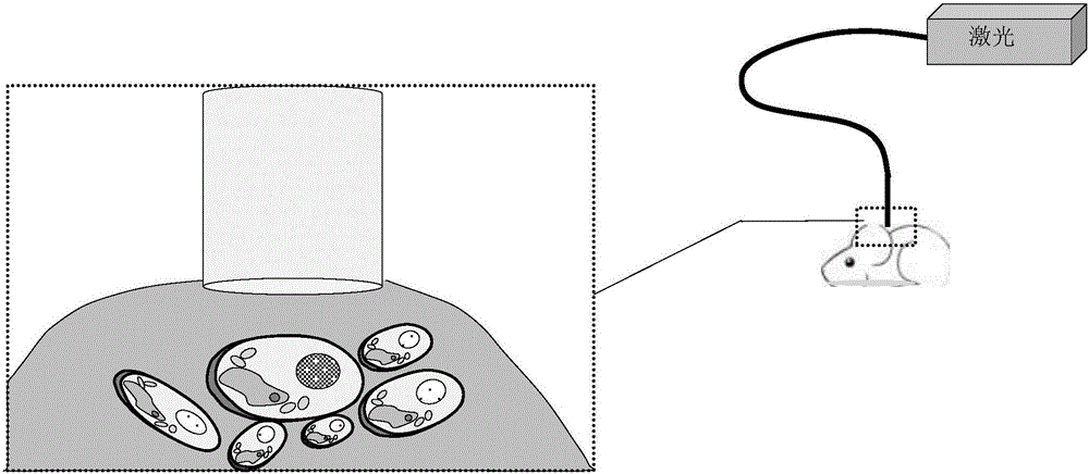
[0031] (4) Take the 1mL cell cryopreservation tube out ...


PUM


Abstract

The invention provides a preparation method and an application method for liquid fluorescent microspheres with a cell tracing function. The preparation method comprises the following steps: adding nano crystal NaYF4:Yb<3+> and Er<3+> into an organic solvent cyclohexane, wherein the cyclohexane can be uniformly dispersed; adding an organic solvent dioctyl phthalate into a mixed solution, wherein the nano crystal is uniformly dispersed in the dioctyl phthalate after cyclohexane is volatilized; by using a capillary injection tube, and injecting the dioctyl phthalate in which the nano crystal is uniformly dispersed into in-vitro culture cells so as to form liquid microspheres in intracellular fluid; injecting cells with the liquid microspheres into a biological body by using a cell syringe. The nano crystal which is uniformly dispersed in the liquid microspheres can emit green fluorescence under the excitation of laser of 980nm, and has a cell tracking function. The invention discloses a completely new in-vivo cell tracing mode which has significant clinical meanings for studying migration, proliferation, metabolism, distribution regulation and the like of cells in bodies.

Description

technical field [0001] The invention relates to a preparation method of biological cell tracer microspheres. The invention also relates to a method for cell tracking of biological cell tracking microspheres. Background technique [0002] Although the traditional cell tracking method can obtain the information of the moment when the animal is sacrificed, it cannot obtain the location, viability, migration, and functional status of the cells in the animal in time, and this method is obviously not conducive to the dynamic observation of the same individual. To address the deficiencies of traditional tracking, researchers have developed different methods for tracking cells in vivo. Currently commonly used tracer techniques include nuclide imaging, magnetic resonance imaging, optical imaging and other techniques. [0003] Up-conversion luminescent nanocrystals are a kind of luminescent material that is excited by low-energy near-infrared light and emits high-energy visible ligh...

Claims


Application Information

Patent Timeline
IPC IPC(8): C09K11/85C09K11/02A61K49/00
CPCA61K49/0017A61K49/0091C09K11/7772
Inventor 杨军王岩刘永军李寒阳
Owner HARBIN ENG UNIV
Why Eureka
Try Eureka