G type enterovirus direct immunofluorescent reagent and kit

A technology of enterovirus and immunofluorescence, which is applied in the direction of disease diagnosis, biological testing, material inspection products, etc., can solve the problems of no detection and diagnosis methods, and achieve the effect of ensuring high sensitivity and high sensitivity

Active Publication Date: 2016-09-28
JILIN UNIV
View PDF3 Cites 5 Cited by
  • Summary
  • Abstract
  • Description
  • Claims
  • Application Information

AI Technical Summary

Problems solved by technology

At present, there is no specific detection and diagnosis method for this type of virus or this type of disease

Method used


Image

  • G type enterovirus direct immunofluorescent reagent and kit
  • G type enterovirus direct immunofluorescent reagent and kit
  • G type enterovirus direct immunofluorescent reagent and kit

Examples


Embodiment 1

[0019] Example 1 Construction of recombinant plasmid for prokaryotic expression of G type enterovirus structural protein VP1

[0020] 1. Primer design

[0021] According to the base composition of the target sequence, design primers for amplification VP1 The upstream and downstream of the gene contain EcoRI and XhoI restriction sites respectively. The primer sequences are as follows:

[0022] Upstream primer: 5' TT GAATTC CAGGCTGGGTATGTGACT 3' (EcoRI)

[0023] Downstream primer: 5'T CTCGAG TTAATGAAAATCACTGAGGGGTT 3' (XhoI)

[0024] 2. Gene amplification

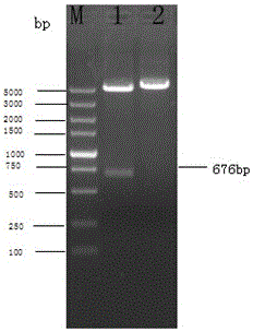
[0025] Genomic RNA of G enteroviruses was extracted by the conventional Trizol method, and cDNA was obtained by using a commercially available conventional reverse transcription kit according to the instructions, and amplified using it as a template VP1 Gene, PCR reaction system:

[0026]

[0027] PCR operating conditions: pre-denaturation at 94°C for 3 min; denaturation at 94°C for 1.5 min, annealing at 54°C for...

Embodiment 2

[0030] Example 2 Expression and purification of VP1 recombinant protein

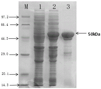
[0031] After the identified positive recombinant plasmid pGEX-4T-1-VP1 was transformed into Escherichia coli BL21(DE3) competent cells, plated and incubated overnight at 37°C, a single colony was picked and inoculated into 3 mL of 100 µg / mL ampicillin containing Cultivate overnight in LB liquid medium, then inoculate 1 mL of the above culture into 200 mL of LB liquid medium containing 100 μg / mL ampicillin and culture at 37 °C until logarithmic growth phase (OD 600 =0.6), adding IPTG (final concentration 1 mmol / L), inducing culture at 20°C for 3 h, and detecting by SDS-PAGE, the recombinant target protein VP1 was obtained, as shown in figure 2 Shown (lane 2). Centrifuge the bacterial cells, and after ultrasonic disruption, wash with 1.5M urea for 3 times to obtain high-purity recombinant protein, such as figure 2 Shown (lane 3).

Embodiment 3

[0032] Example 3 Preparation of monoclonal antibody against G type enterovirus VP1 protein

[0033] 1. Animal immunization: select healthy BALB / c mice aged 6-8 weeks, emulsify the purified VP1 protein with Freund's complete adjuvant, inject 100 µg intraperitoneally into each mouse, and emulsify the protein intraperitoneally with Freund's incomplete adjuvant 14 days later 100µg was injected, and 100µg of purified protein was injected intraperitoneally at the last booster immunization, and 50µg of purified protein was injected into the tail vein 3 to 4 days before fusion.

[0034] 2. Cell fusion: Take splenocytes from immunized mice and mix them with SP2 / 0 in a fusion tube, centrifuge at 300×g for 10 minutes, discard the supernatant, shake the cells to mix the two cells as evenly as possible, and then slowly drop them within 60 seconds Add the preheated PEG-4000 solution slowly to terminate the fusion with serum-free 1640 medium, let it stand still and then centrifuge at 1000 r / ...


PUM


Abstract

The invention discloses a G type enterovirus detecting immunofluorescent reagent and a G type enterovirus immunofluorescent detecting kit. The immunofluorescent reagent is a VP1 monoclonal antibody capable of being labeled with a fluorescent dye, the VP1 monoclonal antibody is obtained by utilizing VP1 protein of the G type enterovirus and adopting a hybridoma monoclonal antibody preparation technology, and the amino acid sequence of the VP1 protein is shown as the sequence table SEQ ID NO.2. The immunofluorescent reagent and the detecting kit can be used for rapid diagnosis of G type enterovirus infection in clinic, and can also be used for localization and identification of a G type enterovirus pathogen in tissue; the direct immunofluorescent detecting time is short, and report results can be generated in 40 min; the specificity is strong, and reaction can only be conducted with the G type enterovirus; the repeatability is good, the accuracy is high, the coincidence rate to an indirect immunofluorescence method can reach 90% or above, and the immunofluorescent reagent and the detecting kit can be used for rapid diagnosis of the G type enterovirus infection.

Description

technical field [0001] The invention belongs to the field of biological technology, and specifically relates to a direct immunofluorescent reagent for detecting G enteroviruses and a kit thereof, which is suitable for the diagnosis of G enterovirus infection and the positioning and identification of sheep enterovirus antigens in tissues or cells. Background technique [0002] Goat G species enterovirus (CEV) belongs to the genus Enterovirus in the family Picornaviridae. Sheep enterovirus infection is a new clinical disease mainly characterized by digestive tract and respiratory tract at home and abroad, which poses a serious threat to the sheep industry. In 2014, the applicant isolated a G type of enterovirus CEV-JL14 from goats in a clinically unknown disease characterized by severe diarrhea, high morbidity and high lethality that broke out in many places in Jilin, and sequenced it And analysis found that the isolated strain is a new enterovirus. At present, there is no s...

Claims


Application Information

Patent Timeline
Patent Type & Authority Applications(China)
IPC IPC(8): G01N33/577G01N33/569G01N33/68
CPCG01N33/56983G01N33/577G01N33/68G01N2333/085G01N2800/065
Inventor 王新平鲁海冰郭昌明王明月刘亚静张群朱利塞王芳
Owner JILIN UNIV
Why Eureka
Try Eureka