
Three-dimensional biomarker detection device, preparation method and method for detecting biomarker

A biomarker and three-dimensional biological technology, applied in the field of three-dimensional biomarker detection devices, can solve the problems of lower detection sensitivity and unreliable detection results, and achieve the effects of improving light transmission, reducing the number of times, and improving quality

Active Publication Date: 2016-11-23
TSINGHUA UNIV
View PDF7 Cites 6 Cited by
  • Summary
  • Abstract
  • Description
  • Claims
  • Application Information

AI Technical Summary

Problems solved by technology

In addition, due to the high water content of the hydrogel, the sample will be diluted when it is dropped on the hydrogel, which not only reduces the sensitivity of the detection, but also may cause unreliability of the detection results.

Method used


Image

  • Three-dimensional biomarker detection device, preparation method and method for detecting biomarker
  • Three-dimensional biomarker detection device, preparation method and method for detecting biomarker
  • Three-dimensional biomarker detection device, preparation method and method for detecting biomarker

Examples


Embodiment 1

[0080] 1. The preparation process of the three-dimensional porous scaffold chip is as follows:

[0081] (1) Preparation of three-dimensional porous scaffold chip

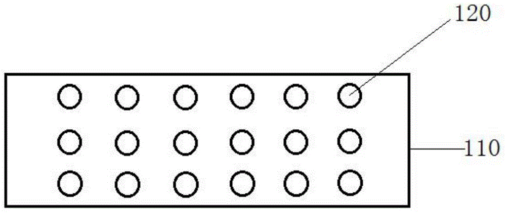
[0082] 1g of polyethylene glycol diacrylate 4000, 0.1g of photoinitiator IG2959, and 0.006g of N-acryloxysuccinimide were dissolved in 6mL of glycerin and 4mL of water to prepare a premix. A 75mm×25mm×1mm glass slide modified with octadecyltrichlorosilane (OTS) and modified with methacryloxypropyltrimethoxy Use a pipette to fill the gap between the two glass slides with the premix solution. Cover the photomask with 6 columns x 16 rows and an aperture of 1.4mm (that is, except for 6 columns and 16 rows of 96 transparent areas with a diameter of 1.4mm, the others are all black opaque plastic films of 75mm x 25mm) On a glass slide, the premix was cross-linked by ultraviolet light to form a cylindrical hydrogel with a diameter of 1.4 mm and a height of 350 microns. After removing the OTS-modified slide and coverslip,...

Embodiment 2

[0092] A three-dimensional biomarker detection device was prepared in the same manner as in Example 1, except that polyethylene glycol, photoinitiator, glycerin, and water were used as raw materials for forming a three-dimensional porous scaffold, and OTS was used for forming a hydrophobic material.

Embodiment 3

[0094] Except that poly(N-isopropylacrylamide), photoinitiator, glycerol, and water were selected as raw materials for forming a three-dimensional porous scaffold, and PDMS was selected as a hydrophobic material, a three-dimensional biomarker detection device was prepared in the same manner as in Example 1.


PUM

PropertyMeasurementUnit
Diameteraaaaaaaaaa

Abstract

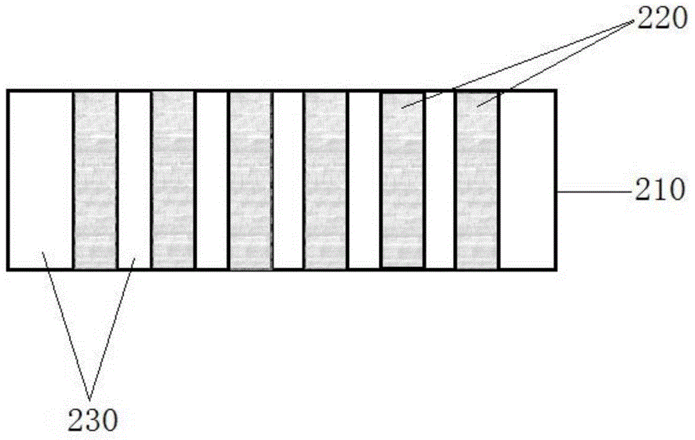
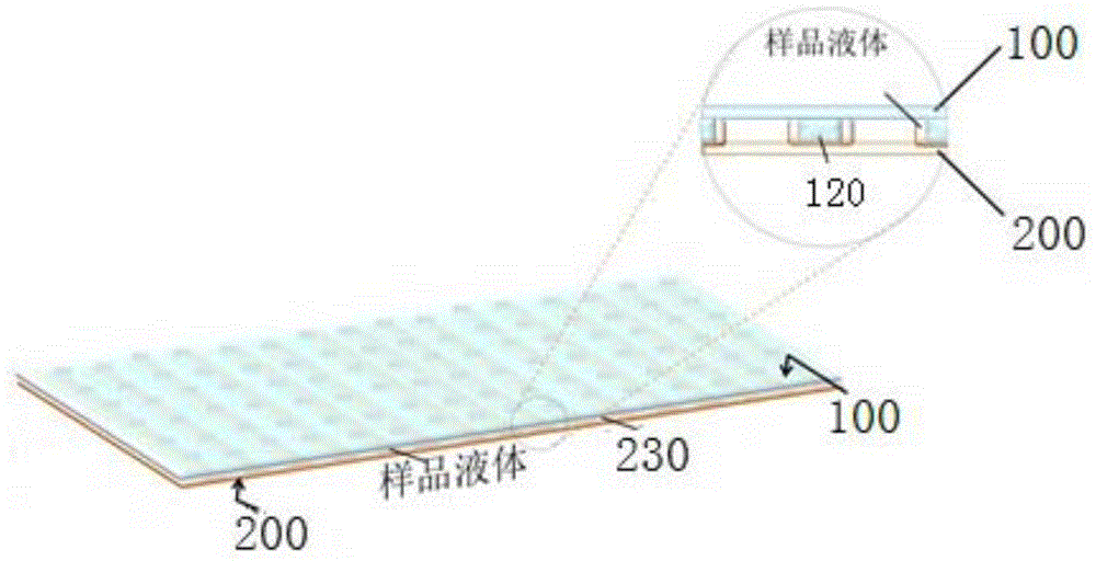
The invention discloses a three-dimensional biomarker detection device, a preparation method and a method for detecting a biomarker. The three-dimensional biomarker detection device includes a three-dimensional porous scaffold chip and a sample carrier plate; wherein the three-dimensional porous scaffold chip comprises a substrate and a plurality of three-dimensional porous scaffolds, the plurality of three-dimensional porous scaffolds are formed on the upper surface of the substrate; the sample carrier plate comprises a base, a plurality of hydrophilic regions and a plurality of hydrophobic regions, the plurality of hydrophilic regions are formed on the upper surface of the base, the plurality of hydrophobic regions are formed on the upper surface of the base, the plurality of hydrophilic regions and the plurality of hydrophobic regions are arranged at intervals, and the plurality of hydrophilic regions are corresponding to the plurality of the three-dimensional porous scaffolds. The three-dimensional biomarker detection device can effectively detect biomarkers.

Description

technical field [0001] The present invention relates to the field of biological detection, in particular to a three-dimensional biomarker detection device, a preparation method of the three-dimensional biomarker detection device and a method for detecting biomarkers using the three-dimensional biomarker detection device. Background technique [0002] The traditional biomarker detection method - ELISA in 96-well plates, is difficult to achieve convenient, low-cost, high-throughput due to its cumbersome experimental steps and large sample volume (50-100 microliters / well). High-volume, high-information detection. The current enzyme-linked immunosorbent assay to increase the amount of information is to fix multiple capture antibodies in the same 96 wells, and the same sample can be added to one well to detect multiple biomarkers at the same time. The anti-catalytic substrate generates luminescent products in situ, and then analyzes the brightness by capturing pictures to simult...

Claims


Application Information

Patent Timeline
IPC IPC(8): G01N33/543
Inventor 杜亚楠鄢晓君
Owner TSINGHUA UNIV
Learn More