
Head thin-layer CT scanning data based frontal bone angle measurement method

A CT scanning and measurement method technology, applied in the fields of medical data mining, image data processing, electrical digital data processing, etc., can solve the problems of decreased measurement accuracy, affecting diagnostic accuracy, and inability to obtain planes, and achieve accurate results. , Easy to operate, and the effect of reducing errors

Inactive Publication Date: 2016-12-21
NANJING CHILDRENS HOSPITAL AFFILIATED TO NANJING MEDICAL UNIV
View PDF3 Cites 0 Cited by
  • Summary
  • Abstract
  • Description
  • Claims
  • Application Information

AI Technical Summary

Problems solved by technology

[0003] At present, the angle of the frontal bone angle is mainly measured based on two-dimensional cranial CT images. This measurement method has the following shortcomings: 1. Radiologists have little understanding of the disease, and different radiologists choose the baseline of CT scans vary, resulting in inaccurate measurements
2. Due to the thick slices of ordinary head CT, the required plane may not be obtained after the film is produced, resulting in measurement errors
The above factors will lead to a decrease in the accuracy of the measurement, which in turn will affect the accuracy of the diagnosis. Therefore, we need to find a measurement method with a higher degree of freedom and better accuracy to improve the prognosis of children with frontal suture early closure. diagnostic accuracy

Method used


Image

  • Head thin-layer CT scanning data based frontal bone angle measurement method
  • Head thin-layer CT scanning data based frontal bone angle measurement method
  • Head thin-layer CT scanning data based frontal bone angle measurement method

Examples


Embodiment 1

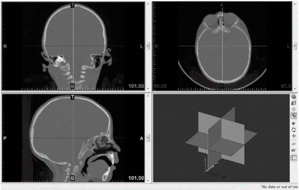
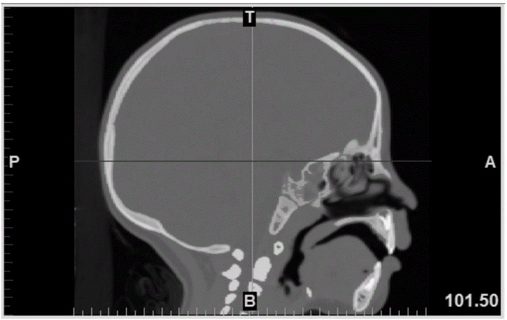
[0023] (1) The patient routinely undergoes thin-slice head CT scanning, and the generated medical digital imaging data is saved in DICOM format and stored in a mobile hard disk.

[0024] (2) Screen the cases that need to be measured.

[0025] (3) Import DICOM data: After opening the mimics software, click the New project wizard button in the toolbar to open the DICOM data screened in step (2), check the information to be imported according to the research needs, and complete the data input.

[0026] (4) Select the comparison scale: use the comparison scale function in the Mimics software, and select BoneScale (bone comparison scale) in the drop-down menu.

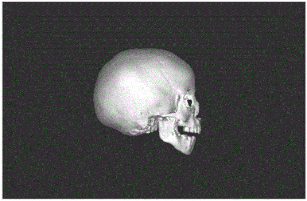
[0027] (5) Create a new mask: create a new mask, and use the threshold selection technology in Mimics software to determine the gray value range: 226Hu to 2976Hu. And use the region growing technology to connect the structure of multiple levels according to the selected gray value, making it a complete head.

[0028] (6) ...


PUM


Abstract

The invention discloses a head thin-layer CT scanning data based frontal bone angle measurement method. According to the invention, head thin-layer CT scanning DICOM image data of a frontal suture premature closure patient is imported by using MINICS software, and a reconstructed three-dimensional image of the head of patient skull is acquired according to the difference of human body bone CT density values. A plane required to be measured is acquired by using methods such as region growing and orthogonal cutting in the MIMICS software, and a frontal bone angle is measured accurately. The method disclosed by the invention is simple in operation and accurate in result, so that a reliable method for measuring the frontal bone angle of frontal suture premature closure child patients is provided, an error caused by human factors such as operations of a doctor in head CT scanning can be significantly reduced, and the method has high accuracy in estimating both the disease severity and surgery effects of the child patients. The frontal bone angle measurement method is simple and convenient to operate and low in time consumption, and does not need to be equipped with professionals, thereby being widely applicable to front-line clinical work.

Description

technical field [0001] The invention belongs to the field of craniofacial plastic surgery, and mainly relates to a method for measuring skull-frontal bone angle after processing thin-layer CT scan data of the skull by using computer three-dimensional reconstruction software (mimics15.0). Background technique [0002] Triangular head deformity, also known as frontal suture syndrome, is a rare type of craniosynostosis, accounting for 7%-23% of craniosynostosis, and its incidence rate accounts for about 1 / 2 of the newborn population. Between 7500 and 1 / 15000, the number and proportion of patients with frontal suture premature closure have increased significantly in recent years. The etiology and pathogenesis of frontal suture syndrome are not yet clear. In the normal population, the frontal suture is the only cranial suture that closes before one year old, and it closes at about 3 to 9 months after birth. Premature suture closure, resulting in frontal ridge protruding from the...

Claims


Application Information

Patent Timeline
IPC IPC(8): G06T7/60G06F19/00G06T17/10
CPCG06T17/10G06T2207/10081G06T2207/30008G16H50/70
Inventor 王刚颜青何俊平高喆钱静祝永杰邱德智郑雷王新法张瑞张献礼丁一
Owner NANJING CHILDRENS HOSPITAL AFFILIATED TO NANJING MEDICAL UNIV
Learn More