Extraction method and purpose of renewable particles of umbilical cord blood

An extraction method and a technology of umbilical cord blood, applied in the fields of biomedicine, histocytology, and regenerative medicine, can solve the problems of immature technology of expanding adult stem cells in vitro and the small number of adult stem cells

Active Publication Date: 2017-02-08
孔五一
View PDF3 Cites 4 Cited by
  • Summary
  • Abstract
  • Description
  • Claims
  • Application Information

AI Technical Summary

Problems solved by technology

[0003] Although stem cells have great potential for clinical treatment, the number of adult stem cells is very small, and the use of stem cells e

Method used


Image

  • Extraction method and purpose of renewable particles of umbilical cord blood
  • Extraction method and purpose of renewable particles of umbilical cord blood
  • Extraction method and purpose of renewable particles of umbilical cord blood

Examples


Embodiment 1

[0069] Example 1. Extraction and cultivation of umbilical cord blood regeneration particles

[0070] In this example, the extraction and cultivation of umbilical cord blood regeneration particles are exemplified.

[0071] Blood source: The umbilical cord blood is collected into a blood bag containing anticoagulant. The anticoagulant is the original sodium citrate in the blood bag, and the umbilical cord blood is transported to company.

[0072] Conditions: The process of obtaining umbilical cord blood regeneration particles requires a complete aseptic operation.

[0073] (1) The umbilical cord blood was taken and centrifuged at 200g for 10 minutes. After centrifugation, it was divided into two parts, the upper layer solution A and the lower layer precipitate A.

[0074] (2) Take the lower layer of precipitate A, add PBS at a volume ratio of 1:2-5 for slight washing, and centrifuge at 200g for 10 minutes. After centrifugation, it is divided into two parts, the upper layer sol...

Embodiment 2

[0080] Example 2. The function of umbilical cord blood regeneration particles to promote the regeneration and repair of myocardial injury

[0081] The experimental group injected the umbilical cord blood regeneration particles (GFP marker) obtained in Example 1 into mice with ischemic myocardial injury through the tail vein, and the control group injected physiological saline. The blood of each mouse was collected every 24 hours after injection for AST detection. After injection, the mouse hearts were collected for 9 consecutive weeks and sectioned to detect the damaged area.

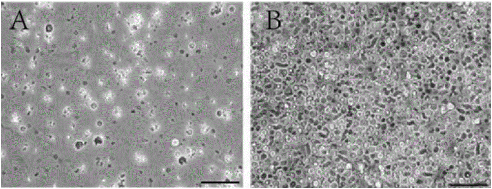
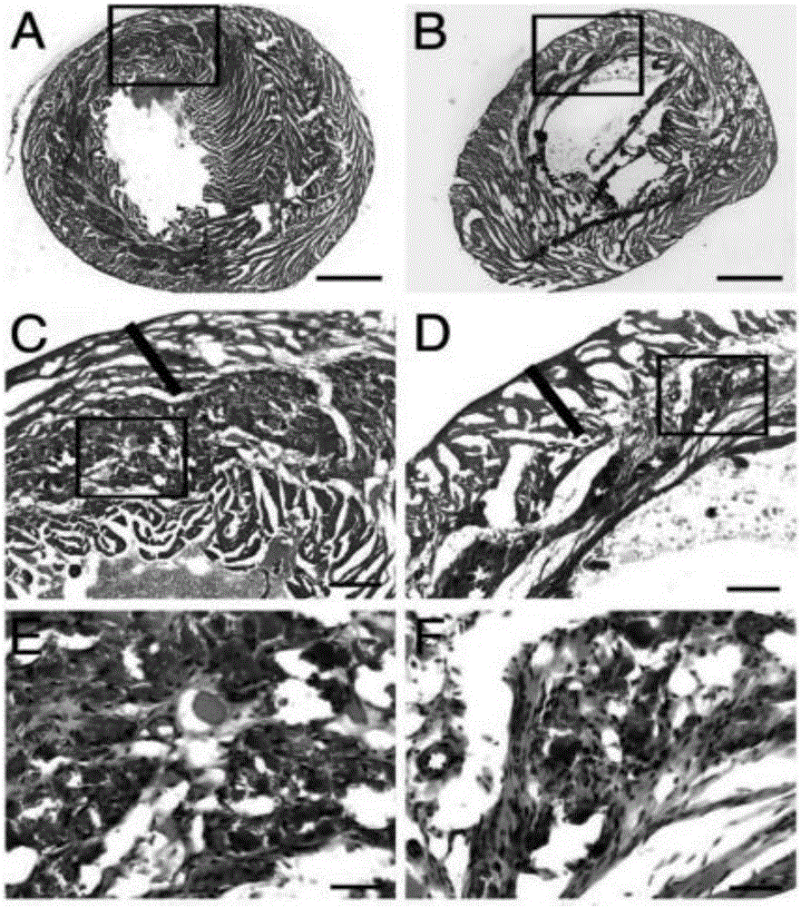
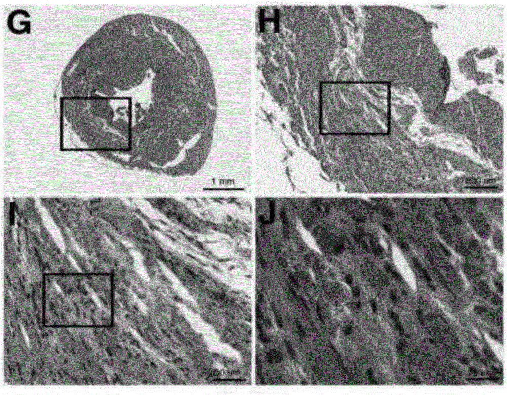
[0082] After transplantation of umbilical cord blood regeneration particles into mice, more cardiospheres accumulated in the myocardial infarction area in the experimental group (see figure 2 , image 3 ), indicating that cord blood regeneration particles formed cardiospheres. Through the detection of Oct4 and CXCR4 in the cardiospheres area of ​​the experimental group (see Figure 4 ), it was foun...

Embodiment 3

[0084] Example 3. The function of umbilical cord blood regeneration particles to promote the regeneration of cerebral blood vessels

[0085] The experimental group injected the regenerative particles (GFP marker) obtained in Example 1 into the mice with ischemic brain injury through the tail vein, and the control group injected normal saline.

[0086] In the 10-week brain examination, it was found that there was no GFP staining in the control group ( Figure 6 A), but there is strong GFP expression in the blood vessel-like structures of the GFP-specific antibody reactive part of the experimental group ( Figure 6 B). Further studies found that GFP and Oct4 co-expressed in blood vessel-like structures ( Figure 7 C-7F) or nestin is also checked ( Figure 7 G-7J).

[0087] The above examples illustrate that the umbilical cord blood regeneration particles of the present invention have the function of promoting the regeneration of brain-injured blood vessels.


PUM


Abstract

The invention provides an extraction method of renewable particles of umbilical cord blood. The extraction method comprises the following steps of taking the umbilical cord blood, carrying out low-speed centrifugation, adding a buffer solution in lower layer precipitates A to carry out washing, carrying out low-speed centrifugation, adding erythrocyte lysate in lower layer precipitates B to carry out cracking, carrying out middle-speed centrifugation, adding the buffer solution in lower layer precipitates C, violently shaking, and carrying out low-speed centrifugation; respectively taking an upper layer solution A, an upper layer solution B, and an upper layer solution D, and carrying out high-speed centrifugation; collecting all the precipitates, and cultivating in an MEM (minimum essential medium); and collecting the medium after being cultivated for 10-30 days, and carrying out 3000-7000g centrifugation so as to enrich the renewable particles of the umbilical cord blood. The renewable particles of the umbilical cord blood can be used for treating ischemic myocardial injury as one of major diseases.

Description

technical field [0001] The invention relates to the field of biomedicine, especially the fields of histocytology and regenerative medicine. It specifically relates to an umbilical cord blood regeneration particle, its extraction method and its application. Background technique [0002] Over the past few decades, medical research related to the treatment of various diseases has developed greatly. Scientists have conducted various studies and discussions, ranging from chemical drugs to gene therapy, and even the currently recognized stem cell therapy. The theory of stem cells was proposed decades ago. In the past ten years, countries all over the world have made important discoveries in the field of stem cells. First, all researchers in the field of stem cells acknowledge that stem cells have a strong regenerative capacity. That is, stem cells have the ability to further differentiate into cells or organs of various tissues in the human body. For example, stem cells can d...

Claims


Application Information

Patent Timeline
IPC IPC(8): A61K35/51A61P9/10A61P9/14A61P17/00A61P1/16A61P13/12
CPCA61K35/51
Inventor 孔五一
Owner 孔五一
Why Eureka
Try Eureka