
Rapid quantitative detection kit for human anti-liver-kidney microsomal antibody

A quantitative detection and kit technology, applied in the field of medicine and biology, can solve the problems of poor flexibility, poor repeatability, and long reaction time.

Active Publication Date: 2017-03-15
ANHUI IPROCOM BIOTECH CO LTD
View PDF7 Cites 0 Cited by
  • Summary
  • Abstract
  • Description
  • Claims
  • Application Information

AI Technical Summary

Problems solved by technology

Membrane immunoassay uses membrane strip chromogenic technology, which is characterized by fixing several items in the same membrane strip for measurement, generally through manual or semi-automatic membrane strip instrument for experimental operation, and finally qualitative judgment by naked eyes, this technology has low sensitivity, The reaction time is long, the detection items can only be fixed and combined, and the flexibility is poor
The sensitivity of enzyme-linked immunosorbent assay has been improved on the basis of membrane strip immunoassay, but it is still low, and the linear range is narrow, the repeatability is poor, and the reaction time is long, which still cannot meet the clinical application well.

Method used


Image

  • Rapid quantitative detection kit for human anti-liver-kidney microsomal antibody

Examples


Embodiment 1

[0025] Embodiment 1 Anti-LKM-1 antibody detection test paper card preparation

[0026] Preparation of nitrocellulose membrane coated with detection line and quality control line:

[0027] Use coating diluent to adjust the concentration of rabbit anti-human CYP2D6 monoclonal antibody (LifeSpan BioSciences Inc.) and goat anti-mouse IgG antibody to 12 mg / ml, and the volume of membrane fluid to 2 μl / cm, and use them as the detection line and quality control respectively The lines were sprayed in parallel on the nitrocellulose membrane for coating, the distance between the test line and the quality control line was 5mm, and then placed in an oven at 37°C for 4 hours. The coating diluent is composed of 100 mM Tris-HCl buffer containing 0.5% BSA and 0.5% sodium lignosulfonate, pH 7.4.

[0028] Preparation of conjugation pads for adsorbing fluorescent microsphere-labeled antigens:

[0029] 1. Aldehydization of rare earth fluorescent microspheres:

[0030] Take 30 mg of rare earth ...

Embodiment 2

[0037] Embodiment 2 test paper card linear range, precision, specificity test and sensitivity test

[0038] Take the anti-LKM-1 antibody standard solution with the content of 0, 5, 20, 50, 100 and 200RU / ml, and measure it with a test paper card.

[0039] Detection method: equilibrate the detection reagent and sample to room temperature, take out the test paper card (prepared in Example 1), and lay it flat; accurately draw 100 μl of standard sample, add it to the sample hole of the test paper card, and analyze it with fluorescent immunochromatography after 20 minutes instrument for testing.

[0040] Linearity: establish a standard curve with the fluorescence intensity value and the corresponding standard substance concentration, specifically Y (fluorescence intensity)=161.1X+17.4, R2 value>0.999, illustrate that the test paper card of the present invention has good linearity in the range of 0-200RU / ml.

[0041] See the table below for the data:

[0042] Anti-LKM-1...

Embodiment 3

[0045] Embodiment 3 stability test

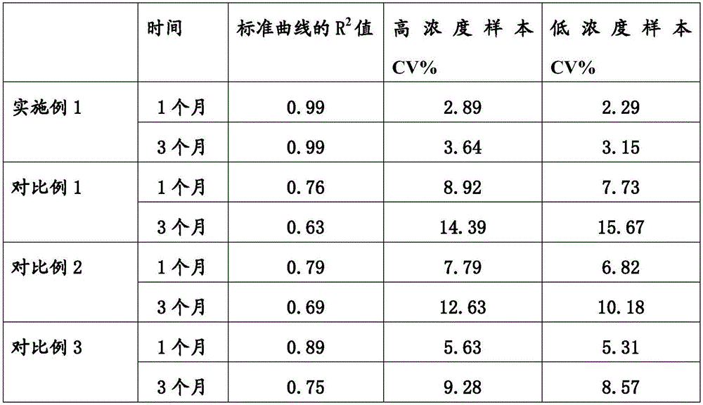
[0046] Place the test paper card prepared in Example 1 in a 37°C environment for 3 months, then determine the standard curve and R of the kit according to the method in Example 2. 2 Value, and adopt the anti-LKM-1 antibody standard substance of 50RU / ml to carry out precision investigation to kit (parallel determination 10 times, calculate CV%), specific result is as follows:

[0047]


PUM

PropertyMeasurementUnit
Diameteraaaaaaaaaa
Sensitivityaaaaaaaaaa

Abstract

The invention relates to a rapid quantitative detection kit for a human anti-liver-kidney microsomal antibody, and belongs to the fields of medical biology. The rapid quantitative detection kit is fluorescence immunochromatographic assay test kit; and the test kit is formed by sequentially mutually overlapping and assembling a treated sample pad, a combination pad with an adsorbed rare earth fluorescent microsphere mark LKM-1 antigen, a nitrocellulose membrane coated with a detection line and a quality control line, and a water absorption pad on a PVC board.

Description

technical field [0001] The invention relates to a rapid quantitative detection kit for human anti-liver-kidney microsome antibody, which belongs to the field of medicine and biology. Background technique [0002] Autoimmune liver diseases (autoimmune liver diseases) mainly include three diseases that are closely related to autoimmunity and mainly focus on liver and gallbladder damage: autoimmune hepatitis (AIH), primary biliary cirrhosis (primary biliary cirrhosis) , PBC) and primary sclerosing cholangitis (primarysclerosing cholangitis, PSC). AIH is a liver disease with circulating autoantibodies and hyperimmunoglobulinemia, unknown etiology, and chronic inflammatory necrosis. PBC is an autoimmune-mediated intrahepatic bile duct injury, followed by liver fibrosis, and eventually leads to hepatic fibrosis. PSC is a chronic syndrome of unknown etiology characterized by chronic cholestasis caused by diffuse inflammation and fibrosis of the extrahepatic and / or intrahepatic bil...

Claims


Application Information

Patent Timeline
IPC IPC(8): G01N33/577G01N33/576G01N33/558G01N33/533G01N21/64
CPCG01N21/6486G01N33/533G01N33/558G01N33/5767G01N33/577
Inventor 陈立国邹伟权张亚丽李庆祥母润红王涛苏焱
Owner ANHUI IPROCOM BIOTECH CO LTD
Learn More