
A kind of EGFR immunohistochemical detection kit based on Fe3O4 nanoprobe and its application method

A detection kit and nanoprobe technology are applied in the field of biological nanomaterials and their preparation to achieve the effects of simple operation, small size and high peroxidase-like activity

Active Publication Date: 2019-04-09
NANJING NANOEAST BIOTECH
View PDF5 Cites 0 Cited by
  • Summary
  • Abstract
  • Description
  • Claims
  • Application Information

AI Technical Summary

Problems solved by technology

[0007] Another object of the present invention is also to provide the core component in the kit of the present invention for detecting the Fe3O4 nanoprobe of EGFR for the defects of existing nanomaterials and biomolecular coupling methods

Method used


Image

  • A kind of EGFR immunohistochemical detection kit based on Fe3O4 nanoprobe and its application method
  • A kind of EGFR immunohistochemical detection kit based on Fe3O4 nanoprobe and its application method
  • A kind of EGFR immunohistochemical detection kit based on Fe3O4 nanoprobe and its application method

Examples


Embodiment 1

[0070] A preparation process of Fe3O4 nanoparticles modified by DMSA:

[0071] Take 28g of FeCl3·6H2O, 80mL of pure water, 20g of FeSO4·7H2O, pass through N2, stir to dissolve completely and heat up to 70°C in a water bath, quickly add 40mL of ammonia water under the action of vigorous stirring. After continuing to stir for 10 min, 10 mL of oleic acid was added dropwise. The reaction was continued at 70 °C for 3 h. Raise the temperature to 90°C to volatilize the ammonia water. After the reaction, it was cooled, magnetically separated, washed twice with ethanol, washed three times with pure water, washed twice with ethanol, dissolved in n-hexane and stored to obtain a n-hexane solution of Fe3O4@OA. Take Fe3O4@OA n-hexane solution (containing 200 mg of Fe3O4@OA nanoparticles), and mix it with an equal volume of DMSA acetone solution (containing 100 mg of DMSA), put it in a three-necked bottle, condense and reflux for 5 hours in a water bath at 60 °C, and wash with pure water f...

Embodiment 2-6

[0073] A preparation process of Fe3O4 nanoprobe in a kit for detecting EGFR expression in human tumor tissue slices based on Fe3O4 nanoprobe:

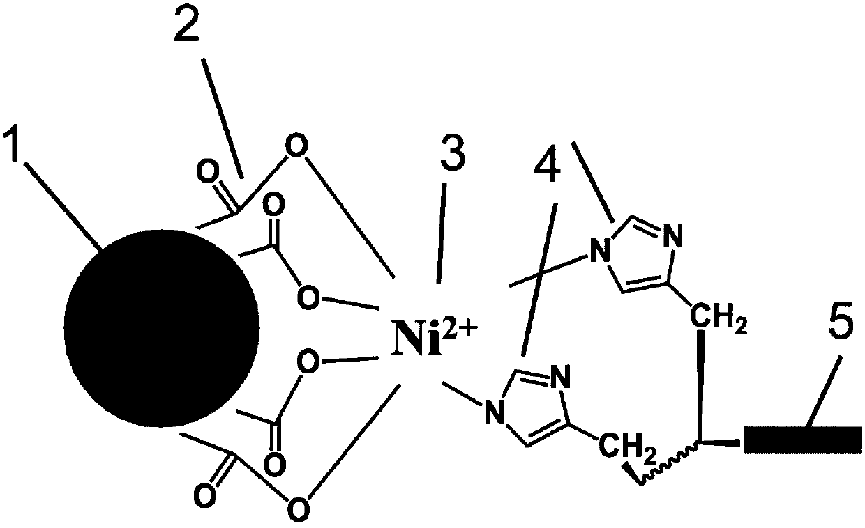
[0074]Get the Fe3O4 nanoparticle (preparation reagent A of Fe3O4 nanoprobe in the kit) 10 μ g obtained in Example 1, and NiSO4 6H2O (Fe3O4 nanoprobe preparation reagent B in the kit) ) were incubated in aqueous solution for 1 h. Centrifuge at a speed of 10000r / min for 20min in a centrifuge to take the precipitate. Resuspend the pellet in 1 mL of pure water, add 50 μg of EGFR single domain antibody (reagent C for Fe3O4 nanoprobe preparation in the kit) and mix well, incubate at 25°C for 30 min, centrifuge at 10,000 r / min for 20 min, and take the precipitate , dissolved in pH 7.4 PBS buffer containing 1% BSA, and stored in a refrigerator at 4°C.

Embodiment 7-15

[0076] A preparation process of Fe3O4 nanoprobe in a kit for detecting EGFR expression in human tumor tissue slices based on Fe3O4 nanoprobe:

[0077] Take 10 μg of Fe3O4 nanoparticles obtained in Example 1 (preparation reagent A of Fe3O4 nanoprobe in the kit), and incubate with 300 μg of NiSO4 6H2O (preparation reagent B of Fe3O4 nanoprobe in the kit) in aqueous solution for 1 h. Centrifuge at a speed of 10000r / min for 20min in a centrifuge to take the precipitate. Resuspend the pellet in 1 mL of pure water, add 10 μg, 15 μg, 20 μg, 25 μg, 30 μg, 35 μg, 40 μg, 45 μg, 50 μg EGFR single domain antibody (reagent C for the preparation of Fe3O4 nanoprobe in the kit) and mix well at 25 Incubate at °C for 30 min, centrifuge at 10,000 r / min for 20 min, take the precipitate, dissolve it with pH 7.4 PBS buffer containing 1% BSA, and store it in a refrigerator at 4°C.


PUM

PropertyMeasurementUnit
sizeaaaaaaaaaa

Abstract

An EGFR immunohistochemical detection kit based on Fe<3>O4 nanoprobes and a using method thereof are disclosed. The kit includes a citric acid restoring powder agent, an endogenous peroxidase blocker, a non-specific binding site blocker, a DAB concentrate liquid, a DAB chromogenic substrate, a DAB diluting liquid, the Fe<3>O4 nanoprobes, a TBS powder agent and a hematoxylin dyeing liquor. A novel method of bonding nanometer particles and an antibody is provided to skillfully combine high peroxidase-like activity of the Fe<3>O4 nanometer particles and biological characteristics of an EGFR single-domain antibody, and a high amount of the EGFR single-domain antibody is loaded on the surfaces of the prepared Fe<3>O4 nanoprobes. The Fe<3>O4 nanoprobes are formed by the Fe<3>O4 nanometer particles surfaces of which are loaded with metal ions and the EGFR single-domain antibody the C end of which is provided with His-Tags, and has advantages of a small size, good stability, high specificity, high sensitivity and high catalytic activity. The EGFR expression level of a tumor pathological tissue can be rapidly and conveniently detected through the kit, thus providing bases for tumor treatment.

Description

technical field [0001] The invention relates to a bionano material and a preparation method thereof, in particular to a Fe3O4 nanometer probe for detecting EGFR and a preparation method thereof. Background technique [0002] Immunohistochemical detection, referred to as immunohistochemical detection, is a specific antibody labeled with a chromogenic agent in situ through antigen-antibody reaction and marker display reaction in tissue cells to locate, characterize, and Bioassay techniques for quantitative determination. Immunohistochemistry is a pathological diagnostic method for tumor diagnosis and differentiation, which is widely used in clinic. Its application directions include: (1) Diagnosis and identification of malignant tumors; (2) Pathological typing of certain types of tumors; (3) Determining the primary site of metastatic malignant tumors; (4) Diagnosis of soft tissue tumors; (5) Identify and discover micrometastases, and provide reference for the formulation of ...

Claims


Application Information

Patent Timeline
Patent Type & Authority Patents(China)
IPC IPC(8): G01N21/78
CPCG01N21/78
Inventor 张宇张薇董金来宋丽娜
Owner NANJING NANOEAST BIOTECH
Learn More