
In-vitro three-dimensional culture method for hepatic cell

A cell, three-dimensional technology, applied in the field of in vitro three-dimensional culture of hepatocytes, can solve the problems of long cell spheroidization time, complex procedures, and high cost of use, and achieve the effects of good biocompatibility, simplified workflow, and extended service life.

Active Publication Date: 2017-04-05
ZHUJIANG HOSPITAL SOUTHERN MEDICAL UNIV
View PDF4 Cites 6 Cited by
  • Summary
  • Abstract
  • Description
  • Claims
  • Application Information

AI Technical Summary

Problems solved by technology

Among them, the suspension spinner bottle culture takes a long time for the cells to form spheres and the rate of spheres is low, and the obtained cell spheres have different sizes and apertures. At the same time, the instrument cannot accurately simulate the physiological state of the cells during the shaking process; by adding polymer substances, the cells can reach Aggregation state, but there are gaps between cells, so that there are defects in the communication between cells. At the same time, if the diameter of the obtained cell sphere is too large, there will be cell necrosis in the center of the cell sphere; the use of photoresist technology is high, and the price Expensive, complicated process, etc.

Method used


Image

  • In-vitro three-dimensional culture method for hepatic cell
  • In-vitro three-dimensional culture method for hepatic cell
  • In-vitro three-dimensional culture method for hepatic cell

Examples


Embodiment 1

[0060] cell culture medium

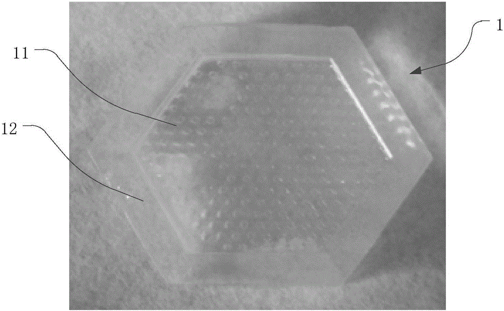
[0061] refer to figure 1 , the cell agglomerate culture matrix 1 of the present invention provides an outer matrix for exosome growth for animal cell culture, and the cell agglomerate matrix 1 comprises a culture area 11 and a fence 12 connected to the periphery of the culture area 11, the enclosure The end face of the baffle 12 is higher than the end face of the culture area 11 to ensure that a certain volume of culture fluid can be accommodated in the culture area 11 . Preferably, the culture area 11 and the enclosure 12 are integrally formed. Considering biocompatibility, the cell culture medium 1 is suitably prepared with agarose.

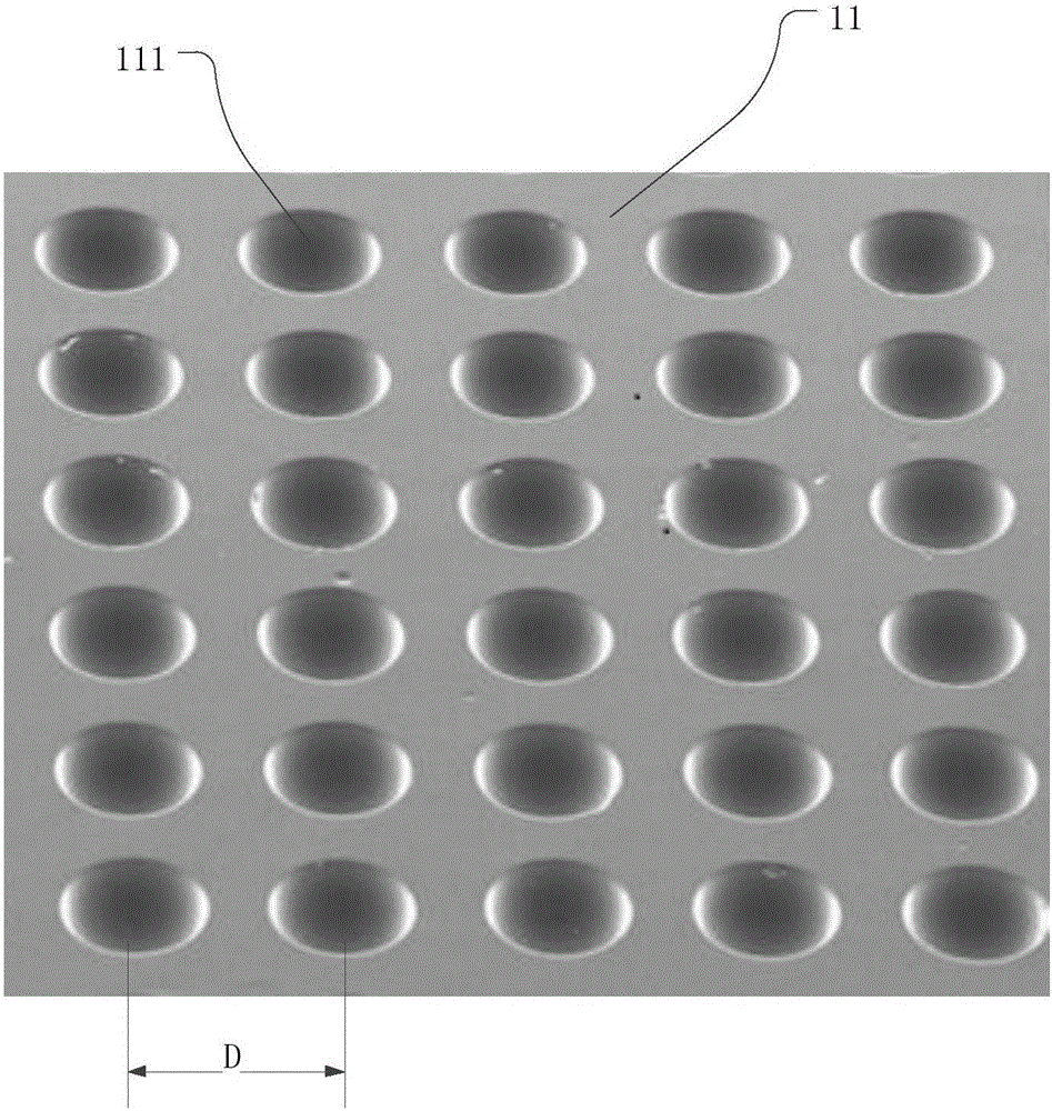
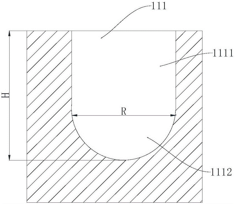
[0062] Further, refer to figure 2 , the culture area 11 is evenly distributed with several polysphere holes 111 for guiding the animal cells growing in the cell polysphere culture substrate 1 to form cell polyspheres, so as to form a three-dimensional structure similar to that in the case of in vivo growth.

[00...

Embodiment 2

[0066] Matrix mold

[0067] refer to Figure 4 , the matrix mold 2 of the present invention is used as a perfusion mold for the cell aggregation sphere culture matrix 1, and the matrix mold 2 includes a first template 21 and a second template 22 nested with each other to form a perfusion molding cavity.

[0068] The first template 21 includes a plate body, a boss 211 disposed inside and a groove 212 circumferentially disposed adjacent to the boss 211 . A number of bumps 213 are evenly distributed on the surface of the boss 211, which are used to correspondingly form aggregated ball holes 111 in the cell aggregated ball culture substrate 1, that is, the shape and size of the outer contour of the bumps 213 are the same as those of the cell aggregated ball culture substrate 1. The internal shape and size of the converging ball holes 111 are the same, and the bumps 213 serve as a positive mold of the converging ball holes 111 .

[0069] Specifically, refer to Figure 5 Each of ...

Embodiment 3

[0080] Physiological indicators of hepatocytes during polysphere culture

[0081] (1) Take human liver cancer cells HepG2, culture them in DMEM high-glucose medium containing 10% fetal bovine serum, penicillin (100u / ml), streptomycin (100u / ml) double antibodies, at 37°C, 5% CO2 , Cultured in an incubator with a relative humidity of 90%, and observed the growth of the cells regularly. Digest with 0.25% trypsin every 2 to 3 days for subculture. After passing to the 5th and 6th generations, take a 24-well plate mold size, inoculate 500 cells per hole in the mold, and culture continuously for 7 days, and measure the relationship between the cell diameter and the number of days of culture.

[0082] like Figure 7 As shown, the cells spread out on the first day of cell inoculation, and the diameter of non-agglomerated balls was measured by Image J software to be about 75um. As the number of days increases, the cells slowly move closer together from the scattered state, the diamet...


PUM


Abstract

The invention discloses a cell sphere gathering culture medium. The cell sphere gathering culture medium comprises a culture area and a fence connected to the periphery of the culture area. The culture area is provided with multiple sphere gathering holes, wherein the lower half portions of the sphere gathering holes are spherical-segment-shaped cavities. The invention further discloses a medium die which comprises a first platy die plate and a second platy die plate, wherein the first platy die plate and the second platy die plate are mutually nested to form a cell sphere gathering culture medium pouring forming cavity. The first die plate is internally provided with a boss and a groove formed adjacent to the boss in the circumferential direction. Multiple protruding points are arranged on the surface of the boss, wherein the upper half portions of the protruding points are spherical segment bodies. A through hole matched with the outer contour of the outer side groove wall of the groove is formed in the second die plate. The invention further discloses a method for conducting animal cell in-vitro three-dimensional sphere gathering culture by utilizing the cell sphere gathering culture medium. The in-vitro three-dimensional culture method for the hepatic cell is relatively low in cost, controllable in scale and simple in process, and the culture effect is greatly superior to that of two-dimensional plate culture.

Description

technical field [0001] The invention relates to an in vitro culture technology of animal cells, in particular to an in vitro three-dimensional culture method of hepatocytes. Background technique [0002] Hepatocytes are widely used as liver substitutes in drug screening, bioartificial livers, and liver transplantation research. Traditional in vitro cell plate two-dimensional culture, on the surface of the hepatocyte growth culture vessel, the cells grow in an adherent island shape, and the cellular protein, urea and drug metabolism functions are not easy to maintain, which are very different from in vivo hepatocytes and cannot replace in vivo hepatocytes. Complete liver function, in order to better simulate the physiological state of liver cells in vivo, increase the interaction between cells and between cells and the extracellular matrix, to enhance the function of cultured liver cells in vitro, and restore the polarity of liver cells. Several in vitro cell three-dimension...

Claims


Application Information

Patent Timeline
IPC IPC(8): C12N5/071
CPCC12N5/0062C12N5/0068C12N5/067C12N2533/76
Inventor 彭青李海燕高毅李阳周树勤张斌斌张志潘明新
Owner ZHUJIANG HOSPITAL SOUTHERN MEDICAL UNIV
Learn More