Pd-l1 fusion protein and use thereof

A fusion protein, PD-L1 technology, applied in the direction of fusion polypeptides, immunoglobulins, peptide/protein components, etc., to achieve the effect of effective treatment

Inactive Publication Date: 2017-04-19
GENEXINE CO LTD +1
View PDF4 Cites 9 Cited by
  • Summary
  • Abstract
  • Description
  • Claims
  • Application Information

AI Technical Summary

Problems solved by technology

However, IgG1 used in conventional Ig fusion techniques can cause antibody-dependent cell-mediated cytotoxicity (ADCC) and complement-dependent cytotoxicity (CDC) in vivo

Method used


Image

  • Pd-l1 fusion protein and use thereof
  • Pd-l1 fusion protein and use thereof
  • Pd-l1 fusion protein and use thereof

Examples


Embodiment 1

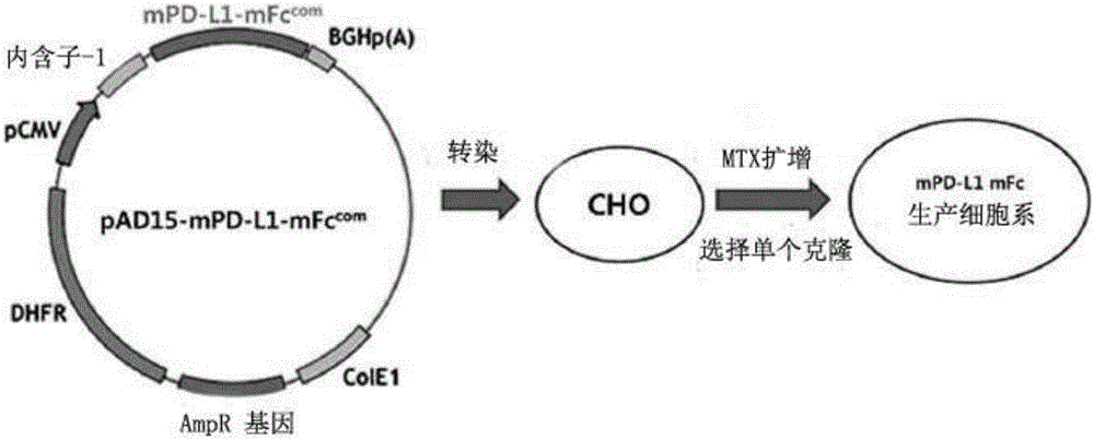
[0170] Example 1: Preparation of mPD-L1-mFc (mouse PD-L1-mouse non-lytic IgG2a) gene construct

[0171] Mouse PD-L1 protein (mPD-L1) has about 70% sequence homology with human PD-L1 protein (hPD-L1). Therefore, when human PD-L1 protein is repeatedly administered to mice, antibodies against human PD-L1 protein (anti-drug antibody, ADA) may be developed, which may be difficult to predict the accurate therapeutic effect of human PD-L1 protein in some cases.

[0172] Therefore, before using the human PD-L1-Fc fusion protein for experiments, the effect of the mouse PD-L1-Fc fusion protein was analyzed by in vivo experiments in mice. A non-soluble Fc (hereinafter referred to as "mFc") was constructed to provide a gene construct capable of producing mPD-L1-mFc, and a cell line expressing mPD-L1-mFc was constructed. The cell line expressing the recombinant protein was cultured in suspension, and the culture medium was collected, after which the recombinant protein was recovered by co...

Embodiment 2

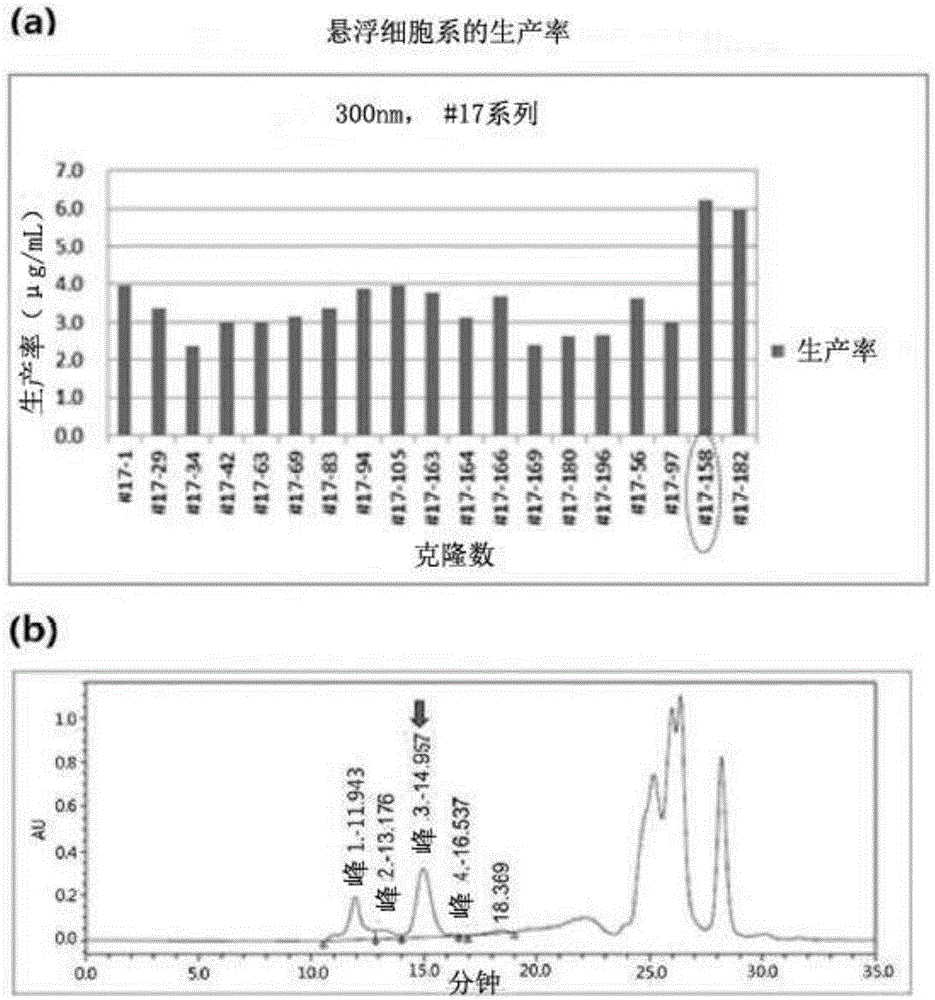
[0175] Example 2: Production of mPD-L1-mFc protein

[0176] In order to mass-produce mPD-L1-mFc (mouse PD-L1-mouse non-lysed IgG2a) protein (SEQ ID NO: 10), cells produced from the mPD-L1-mFc suspension cell line obtained in Example 1 were Isolate and purify target protein from culture medium.

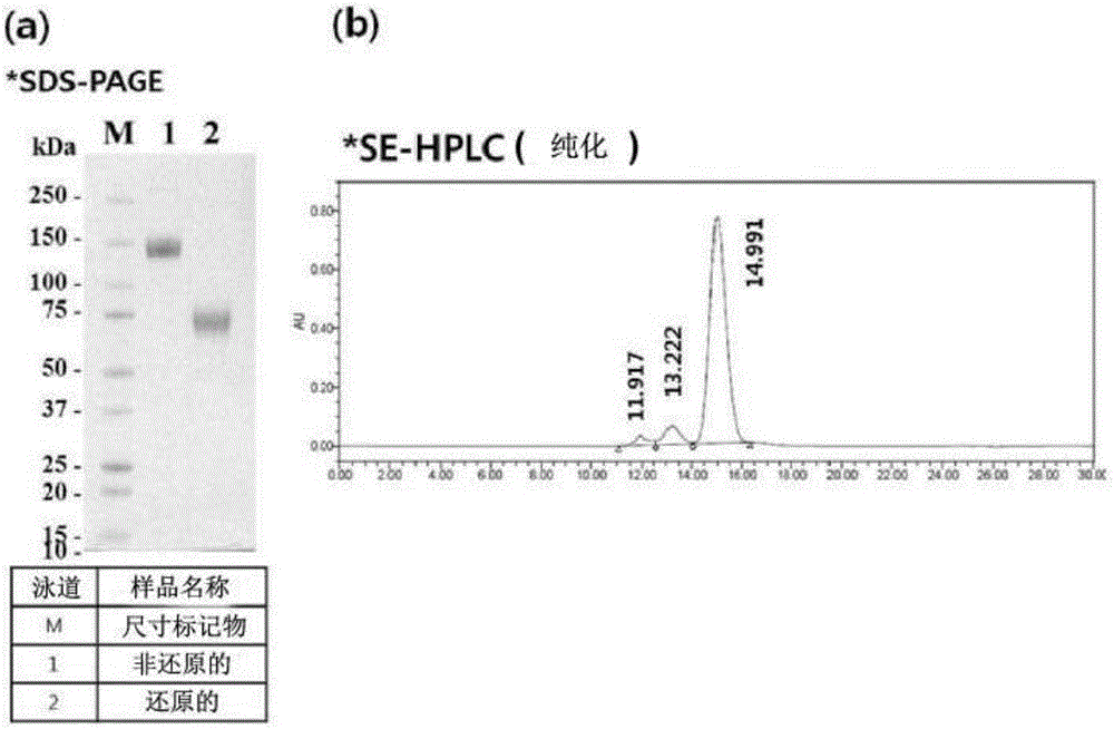
[0177] To purify the protein, the protein purification process was monitored over time at a UV wavelength of 280 nm. As a result, the elution of the target protein was verified by the peak that appeared when the elution buffer (0.1M glycine, pH 3.0) passed through the protein A resin column ( image 3 ). In addition, the purified mPD-L1-mFc protein was analyzed by SDS-PAGE. As a result, the protein size was about 150 KDa under non-reducing conditions and about 75 KDa under reducing conditions, indicating that mPD-L1-mFc is a homodimer form( image 3 a). In addition, the purified product was analyzed by SE-HPLC to confirm its purity ( image 3 b), and measure the level of impurity...

Embodiment 3

[0178] Example 3: Evaluation of in vitro activity of mPD-L1-mFc protein

[0179] In order to evaluate the activity of the purified mPD-L1-mFc protein (SEQ ID NO: 10) in Example 2, the immunosuppressive effect of the protein was analyzed in vitro using mouse splenocytes.

[0180] Such as Figure 4 As indicated, beads were coated with anti-CD3 and mPD-L1-mFc proteins at a ratio of 1:1 or 1:4. In addition, a 10:1 ratio of beads and mouse splenocytes was used to stimulate splenocytes (5x 10 6 Beads: 5x 10 5 spleen cells).

[0181] Mouse splenocytes were stimulated in microwell plates using microbeads coated with anti-CD3 antibody and mPD-L1-mFc protein. After 48 hours, the expression level of cell proliferation factor Ki-67 in mouse splenocytes was analyzed.

[0182] As a result, mPD-L1-mFc protein was observed to inhibit the proliferation of mouse splenocytes (reduced in Ki-67 expression), suggesting that the mPD-L1-mFc fusion protein has immunosuppressive activity ( Figur...


PUM


Abstract

The present invention relates to a fusion protein consisting of an extracellular domain of PD-L1 and a modified immunoglobulin Fc region. The extracellular domain of PD-L1 and a fragment thereof have an excellent immunomodulatory function, and can be used as a drug for immunomodulation when coupled to a modified immunoglobulin Fc region. Accordingly, the PD-L1 fusion protein according to the present invention has been proven to have an excellent effect in inflammatory bowel disease, colitis, psoriasis, asthma, and arthritis disease models, and thus can be used very effectively in treatment of said diseases.

Description

technical field [0001] The present invention relates to a PD-L1 fusion protein prepared by linking PD-L1 to the Fc region of an immunoglobulin, with increased stability and activity. In addition, the present invention relates to a pharmaceutical composition comprising PD-L1 or a specific fragment thereof, and more specifically, to a pharmaceutical composition comprising PD-L1 or a specific fragment thereof for treating immune diseases. Background technique [0002] As a ligand of PD-1 (programmed death-1), human hPD-L1 (human programmed cell death ligand 1) is a type 1 transmembrane protein, which not only in hematopoietic cells such as T lymphocytes, B lymphocytes, It is expressed in dendritic cells or macrophages, but also in non-hematopoietic cells such as keratinocytes, islet cells or hepatocytes. [0003] PD-1 known to bind to PD-L1 is mainly expressed in activated T cells and B cells, macrophages or dendritic cells, and binds to PD-L1 to inhibit cytokine production an...

Claims


Application Information

Patent Timeline
Patent Type & Authority Applications(China)
IPC IPC(8): C07K19/00C12N15/62A61K38/17A61K39/395A61P37/02
CPCC07K14/70596C07K2319/30A61K38/00C07K2317/52A61P1/04A61P17/06A61P19/02A61P29/00A61P37/02A61P37/06A61P3/10A61K38/17A61K39/0005
Inventor 成永哲李智英宋美英林惠星李炳夏
Owner GENEXINE CO LTD
Why Eureka
Try Eureka