Space-dependent type mitochondrial membrane potential fluorescent probe and application thereof

A technology of mitochondrial membrane potential and fluorescent probes, which is applied in the fields of fluorescence/phosphorescence, luminescent materials, and material analysis through optical means. Water solubility and other issues, to achieve good application prospects

Active Publication Date: 2017-04-26
SHANDONG UNIV
View PDF4 Cites 9 Cited by
  • Summary
  • Abstract
  • Description
  • Claims
  • Application Information

AI Technical Summary

Problems solved by technology

However, when the dyeing concentration is slightly low, it is difficult for the two-color fluorescent probe to form aggregates, and when the dyeing concentration is slightly high, it will form a blocky precipitate because of i

Method used


Image

  • Space-dependent type mitochondrial membrane potential fluorescent probe and application thereof
  • Space-dependent type mitochondrial membrane potential fluorescent probe and application thereof
  • Space-dependent type mitochondrial membrane potential fluorescent probe and application thereof

Examples


Embodiment 1

[0028] Synthesis and characterization of LAD

[0029] Add 0.185g of compound 9-(2-ethoxyethyl)-3-formyl-6-(4-vinylpyridyl)-carbazole to a 100mL dry three-necked flask, 25mL of dehydrated methanol, and nitrogen Protect, heat up to 55°C, dissolve after heating for 30min, add 0.168g of compound 6 (N-butyl-4-picoline iodide salt), add 6 drops of piperidine, heat up to reflux, overnight, stop heating, After cooling, the solvent was evaporated to dryness, recrystallized with methanol, and dried to obtain 0.06 g of dark red powder with a yield of about 20%.

[0030] 1H NMR (DMSO-d6, 300MHz): δ (ppm): 8.92 (d, J = 6.6Hz, 2H), 8.55 (m, 4H), 8.24 (t, J = 7.8Hz, 3H), 7.88 (m, 2H), 7.77(dd, J=8.3Hz, 2H), 7.72(s, 1H), 7.58(m, 3H), 7.31(m, 1H), 4.63(t, J=4.8Hz, 2H), 4.49( t,J=7.2Hz,2H).3.78(t,J=6.6Hz,2H),3.38(q,J=7.0Hz,2H),0.91(m,2H),0.32(m,2H),0.95( m,6H).13C NMR(DMSO-d6,75MHz):δ(ppm):153.85,150.49,145.20,144.46,142.90,142.75,141.69,134.30,128.71,127.18,126.88,126.88,126.19,123.01, 12...

Embodiment 2

[0032] Immortalized cell (SiHa and HeLa) and normal cell (HUVEC) culture

[0033] All cell lines were stored at 37°C, 5% CO 2 cultured in a saturated humidity incubator. SiHa and HeLa cell lines were cultured adherently in H-DMEM medium containing 10% fetal bovine serum (containing 1% double antibody). HUVEC cell lines were cultured adherently in M199 medium containing 10% fetal bovine serum (containing 2 ng / mL FGF-2). After the cells grow to the logarithmic phase, culture the slices: ① Soak the coverslips in absolute ethanol for 30 minutes, dry them with an alcohol lamp, and place them in a disposable 35mm culture dish; ② Wash the cells in the 100mL cell bottle with PBS. Three times, digest with 1mL 0.25% trypsin for 3-5 minutes, pour out the medium carefully, add a small amount of fresh medium and pipette evenly, after counting the cells, leave cells with a suitable density, add the medium to the required volume ( The final concentration of control cells was 1×10 5 ), in...

Embodiment 3

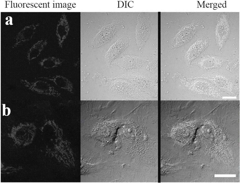
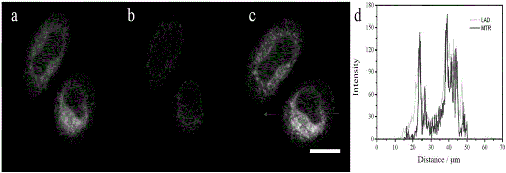
[0035] LAD staining of SiHa and HUVEC cells with normal MMP and counterstaining with MTR

[0036] Wash the inoculated cell slide three times with PBS, stain the cells with 2 μM LAD, and store in CO 2 After incubating in the incubator for 15 minutes, suck out the culture solution and wash it three times with PBS to wash away the unbound excess dye solution. Place the cell growth side down on the glass slide, and observe the stained part of the cell under a laser scanning confocal fluorescence microscope. Fluorescence distribution and brightness changes, etc. It was found that the distribution area of ​​LAD in the cell was filamentous and similar to mitochondria. Therefore, the counterstaining experiment with the commercial mitochondrial probe MTR was used to prove that LAD was in SiHa with normal MMP and SiHa with normal MMP. Whether the staining location in HUVEC cells is mitochondria.

[0037] Wash the inoculated cell slide three times with PBS, stain the cells with 2 μM LAD...


PUM


Abstract

The invention discloses a space-dependent type mitochondrial membrane potential fluorescent probe. The probe is a pyridinium salt compound with a structure as shown in the formula (I). Tests show that the probe can mark internal mitochondria in cells with normal mitochondrial membrane potential in a high selectivity manner and can mark the mitochondria and cell nucleuses in the cells with lowered mitochondrial membrane potential, and the larger the lowering degree of the membrane potential is, the higher the fluorescence intensity of the cell nucleuses is; the probe is targeted to the cell nucleuses after the mitochondrial membrane potential disappears completely. In addition, the probe can also distinguish live and dead cells at the same time, the probe is evidently better than currently-used double-dye related products, and the probe is promising in application prospect in terms of biological researches and medical diagnosis.

Description

technical field [0001] The present invention relates to a fluorescent probe for mitochondrial membrane potential and its application, in particular to a space-dependent fluorescent probe for mitochondrial membrane potential and its ability to display different states of mitochondrial membrane potential in fluorescence imaging or simultaneously detect, display or distinguish between life and death applications in cells. Background technique [0002] Mitochondrial membrane potential (MMP) is an important parameter to represent mitochondrial function, and its decrease indicates the damage of mitochondrial respiratory chain, which leads to cell dysfunction or even death. Therefore, feedback from MMPs in different states is very important for biological research and diagnosis of related diseases. Generally speaking, MMP in healthy cells is normal; MMP in a reduced state indicates some functional disorder, for example, a decrease in sperm MMP indicates a decrease in sperm motilit...

Claims


Application Information

Patent Timeline
IPC IPC(8): C09K11/06C07D401/14G01N21/64
CPCC07D401/14C09K11/06C09K2211/1029G01N21/6486
Inventor 于晓强李学晨孙渝明田明刚
Owner SHANDONG UNIV
Why Eureka
Try Eureka