Multimode MRI longitudinal data-based brain tumor space-time coordinative segmentation method

A collaborative segmentation and multi-modal technology, applied in the field of image processing and biomedicine, can solve the problems of image segmentation with less tumor recurrence, lack of utilization of longitudinal data, and failure to consider the influence of side tissues, so as to improve accuracy and improve Efficiency and the effect of improving segmentation accuracy

Active Publication Date: 2017-05-10
WENZHOU MEDICAL UNIV
View PDF5 Cites 9 Cited by
  • Summary
  • Abstract
  • Description
  • Claims
  • Application Information

AI Technical Summary

Problems solved by technology

[0008] However, most of the existing technologies are based on the segmentation of tumors before surgery, and few images of postoperative and tumor recurrence are segmented, and there is a lack of consideration of using longitudinal data, and the control information of time series images is not fully utilized; In addition, for tumor segmentation methods based on non-longitudinal data, there is a lack of reasonable use of sample point structure information
Although many methods can distinguish the ischemic part from the enhanced part according to the preoperative training image data and do not need to achieve registration, they are actually based on the assumption that the residual tumor part after the operation is the same as before the operation, and the formation of the actual cavity is not considered. The impact on the side tissue does not take into account the factors that cause changes in the void caused by time changes

Method used


Image

  • Multimode MRI longitudinal data-based brain tumor space-time coordinative segmentation method
  • Multimode MRI longitudinal data-based brain tumor space-time coordinative segmentation method
  • Multimode MRI longitudinal data-based brain tumor space-time coordinative segmentation method

Examples


Embodiment Construction

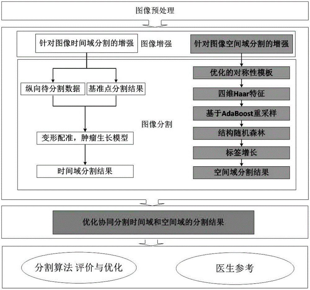
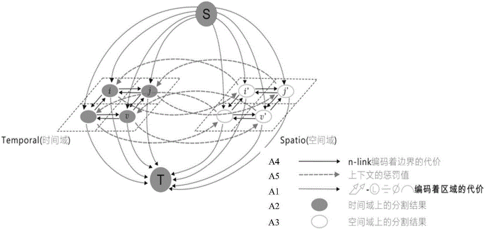
[0030] Specific embodiments of the present invention such as Figure 1-2 Shown is a brain tumor spatio-temporal collaborative segmentation method based on multi-modal MRI longitudinal data, including pre-operative segmentation processing and post-operative segmentation processing. Pre-operative segmentation processing includes the following: obtaining MRI data before brain tumor surgery, and preprocessing the data Afterwards, the pre-operative segmentation results are obtained through the spatial domain segmentation algorithm, and the post-operative segmentation processing includes the following steps: (1) Obtain the MRI data after brain tumor surgery, and preprocess the data; (2) divide the longitudinal data in step (1) into Mapping to the time domain and the space domain for segmentation processing, (3) comparing the time domain segmentation results and the space domain segmentation results to construct a four-dimensional graph model.

[0031] Segmentation is performed accor...


PUM


Abstract

The invention discloses a multimode MRI (Magnetic Resonance Imaging) longitudinal data-based brain tumor space-time coordinative segmentation method. The method comprises pre-operation segmentation processing and post-operation segmentation processing. The post-operation segmentation processing comprises the processes of (1) obtaining brain tumor post-operation MRI data; (2) mapping the longitudinal data to a time domain and a space domain for performing segmentation processing, wherein the time domain segmentation comprises the processes of obtaining a pre-operation segmentation result and longitudinal to-be-segmented data, performing pre-operation and post-operation image registration, and building a post-operation tumor growth model; the space domain segmentation comprises the processes of constructing symmetric templates of different tissues of normal brain, extracting Haar structure features, obtaining an initial probability result by combining a structure random forest method with an AdaBoost framework, performing label growth by utilizing a similar region growth algorithm, and obtaining a space domain segmentation result; and (3) building a four-dimensional graph model in combination with time domain and space domain segmentation results, and optimizing obtained parameters to form an automatic segmentation result. Therefore, the accuracy of brain tumor region segmentation is improved.

Description

technical field [0001] The invention belongs to the technical field of combining image processing and biomedicine, in particular to a space-time collaborative segmentation method for brain tumors based on multimodal MRI longitudinal data. Background technique [0002] Brain tumors (brain tumors) are a serious threat to human health. Magnetic Resonance Imaging (MRI, Magnetic Resonance Imaging) segmentation is an important prerequisite for brain tumor surgery radiotherapy planning and long-term longitudinal research. The present method is of great importance for brain tumor segmentation based on multimodal MRI longitudinal data. Brain tumors refer to cancerous substances growing in the cranial cavity, including primary tumors caused by lesions in the brain parenchyma, and secondary tumors that metastasize and invade the brain from other parts of the body. It has a high incidence in the population and has become one of the important tumors that endanger people's life and heal...

Claims


Application Information

Patent Timeline
Patent Type & Authority Applications(China)
IPC IPC(8): G06T7/11G06T7/194G06K9/62
CPCG06T2207/30016G06T2207/30096G06T2207/10088G06F18/2148
Inventor 潘志方叶夏陈峰
Owner WENZHOU MEDICAL UNIV
Why Eureka
Try Eureka