
Hepatocellular carcinoma marker

A technology of hepatocellular carcinoma and markers, applied in biological testing, biomaterial analysis, instruments, etc., can solve problems such as stagnation and failure to provide

Active Publication Date: 2017-05-10
NAT INST OF ADVANCED IND SCI & TECH
View PDF22 Cites 5 Cited by
  • Summary
  • Abstract
  • Description
  • Claims
  • Application Information

AI Technical Summary

Problems solved by technology

As a result, a variety of hepatocellular carcinoma markers have been identified, and some achievements have been made in the field of pathological analysis of liver diseases. Excellent markers of AFP (particularly the AFP-L3 fraction) used in the clinic, now stagnant

Method used


Image

  • Hepatocellular carcinoma marker
  • Hepatocellular carcinoma marker
  • Hepatocellular carcinoma marker

Examples


preparation example Construction

[0246] (1) 18 Preparation of O-labeled peptides

[0247] The culture supernatants of two kinds of hepatocellular carcinoma culture strains (HLF strain and HAK1A strain) and protein samples prepared from the cancerous and non-cancerous parts of the pathological tissues of hepatocellular carcinoma patients were injected into the NPA lectin-bound The column captures the NPA-binding glycopeptide group, treats it with trypsin and fragments it into peptides, and passes through the NPA lectin column again to capture the NPA lectin-binding glycopeptide group again. The obtained candidate glycopeptides are treated with peptide-N-glycosidase (glycopeptidase F, PNGase) to remove the N-binding sugar chains and replace them with the introduction of Asn with sugar chains 18 O to label the peptide with a stable isotope. (Thus, whether or not a sugar chain is bound to the peptide was clearly confirmed by experiments, and to which Asn on the peptide sequence the sugar chain was bound was kno...

Embodiment 1

[0321] (Example 1) Tissue lectin array resolution

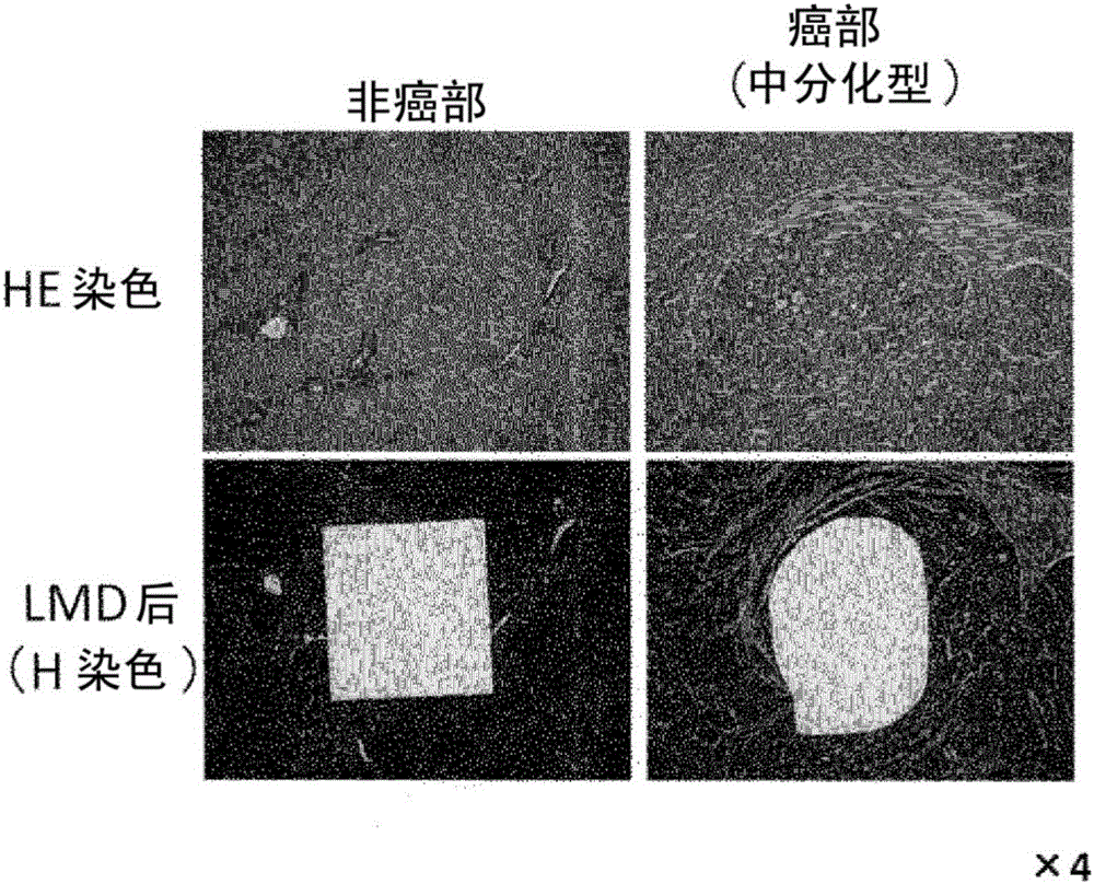
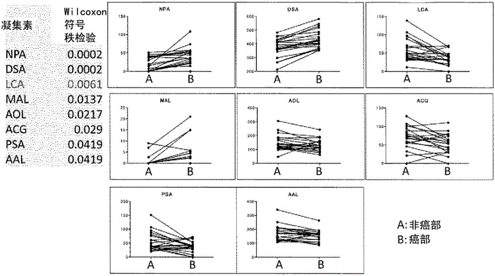
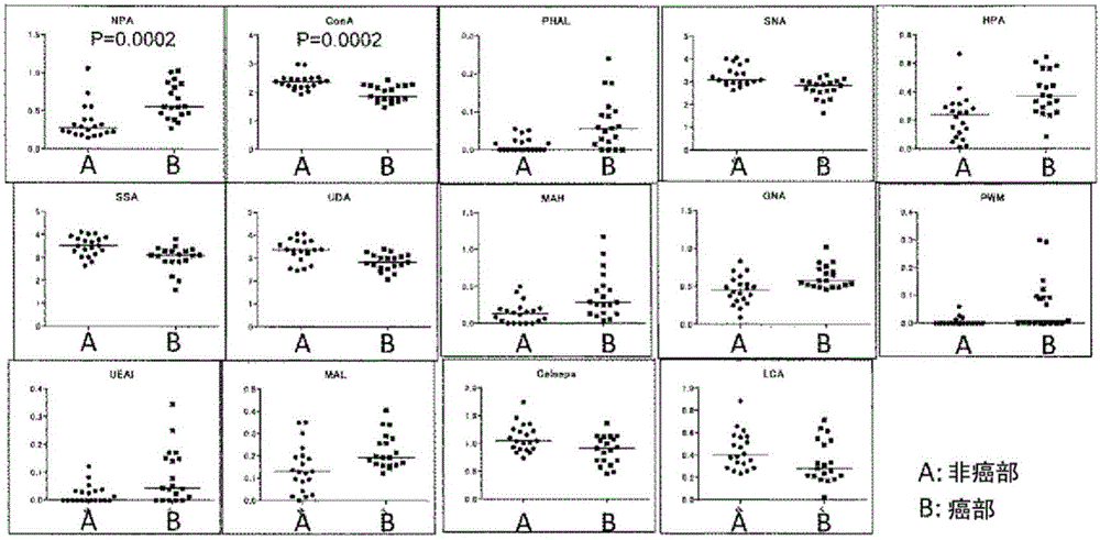
[0322] The lectin microarray used in this study is a system that immobilizes 45 kinds of plant lectins with different specificities on the same substrate, and simultaneously analyzes the interactions (binding properties) with sugar chains on glycoproteins to be analyzed (Kuno et al., Nature Methods 2, 851-856, 2005). Using this system, an attempt was made to screen for a lectin that forms a significant high signal value in a quantitative assay system for hepatocellular carcinoma tissue, or an optimal lectin that can specifically stain cancer in tissue staining. In this experiment, formalin-fixed and paraffin-embedded liver tissues from patients with HCC were used. Specific areas of cancerous and non-tumor liver parenchyma (non-cancerous) were made into tissue fragments by laser microdissection (LMD) and collected, followed by protein extraction and lectin array analysis after fluorescent labeling. The basic protocol follo...

Embodiment 2

[0338] (Example 2) NPA lectin reaction of hepatocellular carcinoma cultured cells and liver cancer patient tissues based on sandwich ELISA Responsive research

[0339] Seven cultured hepatocellular carcinoma cell lines (HuH-7, HepG2, KYN-1, KYN-2, HAK-1A, HAK-1B, HLF) whose reactivity to NPA was previously confirmed by lectin array were studied and Can Liver Cancer Patient Tissues Be Constructed? Figure 5 The sandwich ELISA system shown in b. It should be noted that the basic protocol for extracting protein from cultured cells to fluorescent labeling was carried out according to the method of Tateno et al. or Toyoda et al. (Methods Enzymol 478, 181-195, 2010, Genes Cells 16, 1-11, 2011). It should be noted that the preparation of samples from tissue specimens was carried out according to Example 1.

[0340] (2-1) Protein extraction from cultured cells

[0341] Adjust the cultured liver cancer cell lines to 2×10 in each 1.5mL tube 6 indivual. After the pellets were fo...


PUM

PropertyMeasurementUnit
thicknessaaaaaaaaaa

Abstract

The present invention addresses the problem of providing a hepatocellular carcinoma marker which can be used for detecting the presence of hepatocellular carcinoma and comprises a glycoprotein that can occur in the liver only when the carcinoma is developed regardless of the change in the condition of the liver. The present invention provides a hepatocellular carcinoma marker which comprises an NPA lectin-binding glycoprotein containing an NPA lectin-binding sugar chain epitope having at least one property selected from the following properties (1) to (5) (1) the sugar chain epitope does not contain core fucose (a fucose alpha 1(arrow) 6 sugar chain); (2) the sugar chain epitope contains a composite sugar chain that contains three (less than four) mannose molecules; (3) the sugar chain epitope does not contain a high-mannose-type sugar chain containing five or more mannose molecules; (4) the sugar chain epitope comprises a composite sugar chain that does not rely on the bindability to LCA lectin; and (5) the sugar chain epitope comprises a composite sugar chain that does not rely on the bindability to ConA lectin. The presence of the development of hepatocellular carcinoma or the degree of the progression or malignancy of the carcinoma can be determined by detecting the hepatocellular carcinoma marker of the present invention in a sample of interest.

Description

technical field [0001] The present invention relates to a novel hepatocellular carcinoma marker for accurate and simple diagnosis of hepatocellular carcinoma, and a method for examining hepatocellular carcinoma using the marker. More specifically, it relates to an examination method for early detection of hepatocellular carcinoma and prediction of the prognosis of a patient suffering from cancer, and further relates to an examination kit for the examination. Specifically, glycoproteins that are not expressed in non-cancerous regions of liver tissue but specifically expressed in hepatocellular carcinoma in cancerous regions or in the mesenchymal site (TME) surrounding cancer cells are identified, thereby providing Protein markers of hepatocellular carcinoma. In addition, it relates to providing a detection method of hepatocellular carcinoma using a lectin that binds to the glycoprotein and a kit used in the detection method. Background technique [0002] In Japan, cancer (m...

Claims


Application Information

Patent Timeline
Patent Type & Authority Applications(China)
IPC IPC(8): G01N33/574G01N1/30G01N33/48G01N33/53G01N33/543
CPCG01N1/30G01N2400/02G01N33/57438G01N33/57492G01N2333/71G01N2333/70596G01N2333/96472
Inventor 久野敦佐藤隆松田厚志成松久梶裕之栂谷内晶调宪前原喜彦
Owner NAT INST OF ADVANCED IND SCI & TECH
Learn More