Isolated culture method for cartilage cells

A technology of separation culture and cell culture, applied in the field of cell culture, which can solve problems such as insufficiency of digestion, enzyme toxicity damage, difference in cell quantity and quality, and achieve the effect of maintaining vitality and improving efficiency

Inactive Publication Date: 2017-05-24
GUANGZHOU SALIAI STEMCELL SCI & TECH CO LTD
View PDF0 Cites 1 Cited by
  • Summary
  • Abstract
  • Description
  • Claims
  • Application Information

AI Technical Summary

Problems solved by technology

Although the method of mechanical separation of tissue blocks is simple to operate, the cell separation cycle is long, so it is only suitable for studies with a small demand for cells
Compared with the mechanical separation method of tissue blocks, the collagenase digestion met

Method used


Image

  • Isolated culture method for cartilage cells
  • Isolated culture method for cartilage cells

Examples


Embodiment 1

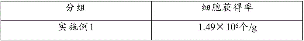
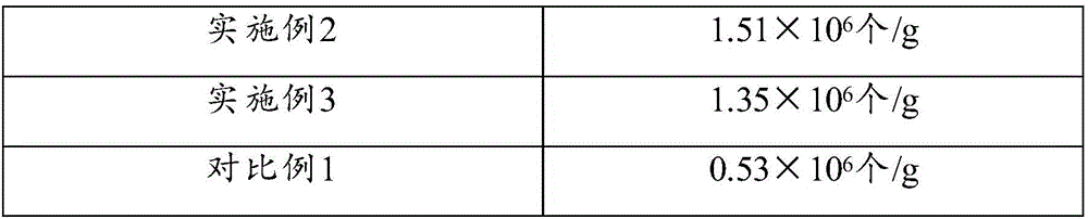
[0027] Example 1: Isolation of Chondrocytes

[0028] 4-week-old New Zealand rabbits were sacrificed by ear vein injection, the articular cartilage of the hindlimb was separated under aseptic conditions, the fascia and perichondrium covering the cartilage tissue were peeled off, and put into a glass petri dish filled with PBS. The isolated cartilage tissue was cut into small pieces smaller than 1 mm.

[0029] The cartilage tissue was digested with 0.25% trypsin for 20 min; after washing with PBS for 3 times, it was digested with 0.05% type II collagenase for 18 h, and the cells were collected every 3 h.

[0030] Count the collected cells and inoculate them on 25cm 2 In cell culture flasks, add 1×10 5 chondrocytes in 5 mL of DMEM / F12 medium containing 10% FBS and 0.1 μg / mL EGF.

Embodiment 2

[0031] Example 2: Isolation of Chondrocytes

[0032] 4-week-old New Zealand rabbits were sacrificed by ear vein injection, the articular cartilage of the hindlimb was separated under aseptic conditions, the fascia and perichondrium covering the cartilage tissue were peeled off, and put into a glass petri dish filled with PBS. The isolated cartilage tissue was cut into small pieces smaller than 1 mm.

[0033] Add 0.25% trypsin to the cartilage tissue to digest for 30 minutes; after washing with PBS for 3 times, enzymatically digest with 0.05% type II collagenase for 18 hours, and collect cells every 3 hours.

[0034] Count the collected cells and inoculate them on 25cm 2 In cell culture flasks, add 1×10 5 chondrocytes in 5 mL of DMEM / F12 medium containing 10% FBS and 0.1 μg / mL EGF.

Embodiment 3

[0035] Example 3: Isolation of Chondrocytes

[0036] 4-week-old New Zealand rabbits were sacrificed by ear vein injection, the articular cartilage of the hindlimb was separated under aseptic conditions, the fascia and perichondrium covering the cartilage tissue were peeled off, and put into a glass petri dish filled with PBS. The isolated cartilage tissue was cut into small pieces smaller than 1 mm.

[0037] The cartilage tissue was digested with 0.25% trypsin for 20 min; after washing with PBS for 3 times, it was digested with 0.1% type II collagenase for 18 h, and the cells were collected every 3 h.

[0038] Count the collected cells and inoculate them on 25cm 2 In cell culture flasks, add 1×10 5 chondrocytes in 5 mL of DMEM / F12 medium containing 10% FBS and 0.1 μg / mL EGF.


PUM


Abstract

The invention relates to the technical field of cell culture, in particular to an isolated culture method for cartilage cells. The isolated culture method comprises the following steps: treating a cartilage tissue into tissue blocks, adding trypsin for digestion, and then adding type II collagenase for digestion, wherein the concentration by mass percentage of the type II collagenase is 0.05 to 0.1 percent, and the digestion time of the type II collagenase is 16 to 20 hours. An experimental research shows that by the adoption of proper concentration and digestion time of type II collagenase for digestion of the cartilage tissue, the vitality of the cartilage cells can be effectively retained, so that enough number of cartilage cells can be obtained within relatively short time during later multiplication culture of the cartilage cells obtained by separation, and the cell culture efficiency is greatly improved.

Description

technical field [0001] The invention relates to the technical field of cell culture, in particular to a method for separating and culturing chondrocytes. Background technique [0002] Articular cartilage is a kind of hyaline cartilage composed of chondrocytes, cartilage matrix and type II collagen. Trauma and inflammation of joints can cause damage to articular cartilage. Damage to the articular cartilage can cause patients to experience recurring joint pain and limited mobility, severely impacting their daily lives. Due to the limited regenerative capacity of articular cartilage, once damaged, it is difficult to repair itself. The repair of articular cartilage damage has always been one of the important topics in orthopedic research. [0003] Cartilage tissue engineering technology is to culture and expand cartilage seed cells in vitro, and plant them at a higher concentration on a scaffold material with good biocompatibility and degradability to construct tissue engineere...

Claims


Application Information

Patent Timeline
IPC IPC(8): C12N5/077
CPCC12N5/0655C12N2509/00
Inventor 陈海佳葛啸虎王一飞赵萌萌王小燕
Owner GUANGZHOU SALIAI STEMCELL SCI & TECH CO LTD
Why Eureka
Try Eureka