
Fluorescence detection method for hydrolase activity

A fluorescence detection and hydrolytic enzyme technology, applied in the field of biological analysis, can solve the problems of insufficient anti-interference ability, poor anti-interference ability and high detection cost, and achieve the effect of short detection time, strong anti-interference ability and high sensitivity detection

Inactive Publication Date: 2017-05-24
NANJING UNIV OF SCI & TECH
View PDF4 Cites 5 Cited by
  • Summary
  • Abstract
  • Description
  • Claims
  • Application Information

AI Technical Summary

Problems solved by technology

Taking alkaline phosphatase as an example, there are two types of fluorescence analysis methods currently available for its activity determination: (1) Based on the principle of polymerization-induced luminescence, alkaline phosphatase hydrolyzes water-soluble phosphoric acid-tetraphenylethylene to form insoluble Tetraphenylethylene in water, tetraphenylethylene will produce fluorescence after polymerization, so as to realize the fluorescence analysis of alkaline phosphatase, but this method has defects such as poor selectivity, low sensitivity and insufficient anti-interference ability (1.Gu X, et al. (2013).A new fluorometric turn-on assay for alkaline phosphatase and inhibitor screening based on aggregation and deaggregation oftetraphenylethylene molecules[J].Analyst,138(8):2427-2431; 2.Liang J,et al.(2013). Fluorescent light-up probe with aggregation-induced emission characteristics for alkaline phosphatase sensing and activity study[J].ACS Applied Materials&Interfaces,5(17):8784-8789.); (2) Using carbon quantum dots as fluorescent probes, carbon quantum dots When the carboxyl groups enriched on the surface are triggered to aggregate, the fluorescence can be quenched, thereby realizing the fluorescence analysis of alkaline phosphatase. However, this method has defects such as high detection cost, poor anti-interference ability and possible toxicity (1. Qian Z, etal.(2015).Carbon quantum dots-based recyclable real-time fluorescence assayfor alkaline phosphatase with adenosine triphosphate as substrate[J].Analytical Chemistry,87(5):2966-2973; 2.Qian Z S,et al .(2015).A real-time fluorescent assay for the detection of alkaline phosphatase activity based on carbon quantum dots[J].Biosensors and Bioelectronics,68:675-680.)

Method used


Image

  • Fluorescence detection method for hydrolase activity
  • Fluorescence detection method for hydrolase activity
  • Fluorescence detection method for hydrolase activity

Examples


Embodiment 1

[0034] The method of the invention is used in a feasibility analysis experiment for detecting alkaline phosphatase activity.

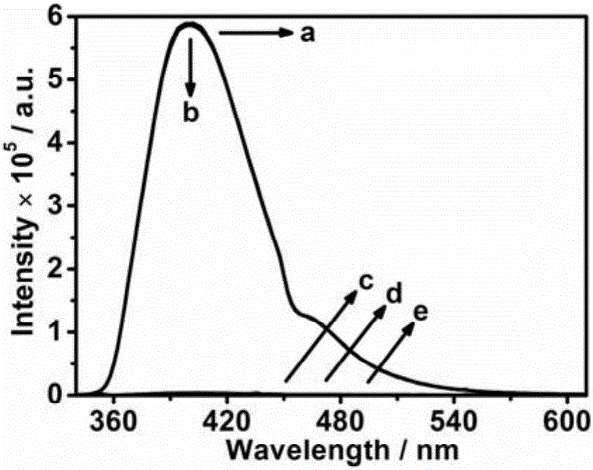
[0035] 20μL 0.05mg mL -1 Alkaline phosphatase samples were added to 2980 μL containing 2 mM ascorbic acid-2-phosphate and 0.03 mM Cu 2+ - After incubating at 25° C. for 15 minutes in the detection solution of the bathocuproine disulfonic acid fluorescent probe, record the fluorescence emission spectrum of the detection solution.

[0036] In feasibility analysis experiments, missing components were replaced with the same volume of buffer. from figure 1 As can be seen, when alkaline phosphatase (a) or substrate (b) are not added in the detection solution, the fluorescence emission intensity of the detection solution at 402nm is stronger; in the detection solution, bathocuproine disulfonic acid ( c), without adding Cu 2+ - When the bathocuproine disulfonic acid fluorescent probe (d) or all components are present (e), the fluorescence emission intensit...

Embodiment 2

[0038] The method of the invention is used to detect the relationship between the fluorescence emission intensity of the alkaline phosphatase activity and the alkaline phosphatase activity.

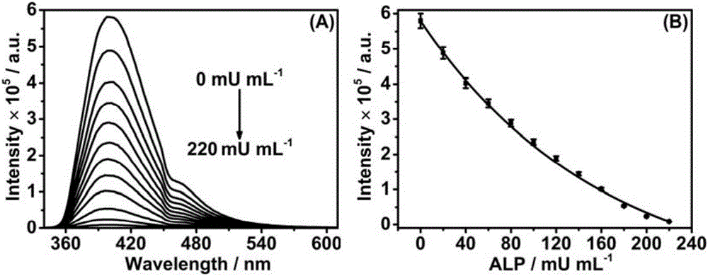
[0039] Make 20 μL of concentrations of 0, 20, 40, 60, 80, 100, 120, 140, 160, 180 and 220 mU mL -1 The alkaline phosphatase samples were added to 2980 μL containing 2 mM ascorbic acid-2-phosphate and 0.03 mM Cu 2+ - After incubating at 25° C. for 15 minutes in the detection solution of the bathocuproine disulfonic acid fluorescent probe, record the fluorescence emission spectrum of the detection solution.

[0040] from figure 2 It can be seen that with the increase of the alkaline phosphatase activity in the sample, the fluorescence emission intensity of the detection solution at ~402nm is correspondingly weakened, and the fluorescence emission intensity has a good quantitative relationship with the alkaline phosphatase activity. It can be seen that the fluorescence analysis method of ...

Embodiment 3

[0042] The method of the invention is used to detect the selectivity of alkaline phosphatase activity.

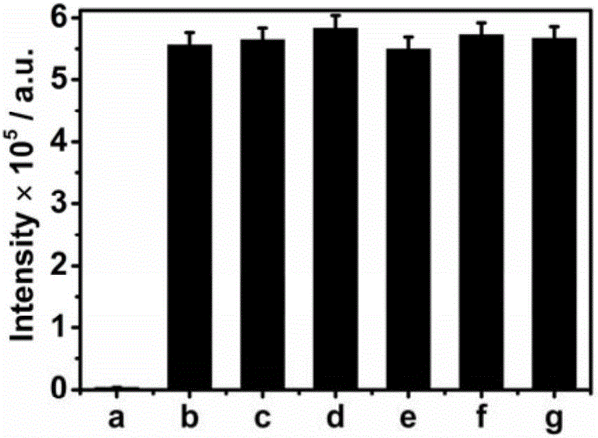
[0043] 20μL 0.05mg mL -1 Alkaline phosphatase or 20 μL 0.5mg mL -1 The other protein solution was added to 2980 μL containing 2 mM ascorbic acid-2-phosphate and 0.03 mM Cu 2+ - In the detection solution of the bathocuproine disulfonic acid fluorescent probe, after incubating at 25° C. for 15 minutes, record the fluorescence emission intensity of the detection solution at ~402 nm.

[0044] from image 3 It can be seen that compared with the blank control, although the concentration of other proteins is 10 times the concentration of alkaline phosphatase, only when alkaline phosphatase is added, the fluorescence emission intensity of the detection solution at ~402nm will decrease significantly. It can be seen that the fluorescence analysis method of the present invention has high selectivity when used to detect alkaline phosphatase activity.


PUM


Abstract

The invention discloses a fluorescence detection method for hydrolase activity. The method comprises the following steps: hatching detection liquid containing a substrate and a Cu<2+>-bathocuproine disulfonic acid complex with a to-be-detected hydrolase solution, taking the Cu<2+>-bathocuproine disulfonic acid complex as a fluorescent probe, and reducing the fluorescent probe by a reducing product obtained by hydrolyzing the substrate by virtue of hydrolase, so as to generate the Cu<2+>-bathocuproine disulfonic acid complex and weaken a fluorescent signal of the detection liquid. The higher is the activity of the hydrolase in a sample, the lower fluorescence emission intensity of the detection liquid is. The fluorescence detection method disclosed by the invention has high detection sensitivity on hydrolase activity, good selectivity, strong interference resistance, good reproducibility, simple operation and short detection time and can be used for quick and high-sensitivity detection of the hydrolase activity in a clinical sample, and the detection cost is greatly reduced.

Description

technical field [0001] The invention belongs to the technical field of biological analysis and relates to a fluorescence detection method for hydrolase activity. Background technique [0002] Hydrolase is a general term for a class of enzymes that catalyze hydrolysis reactions, and widely exists in various animals, plants and microorganisms. Simple, rapid and sensitive detection of hydrolase activity is of great significance for early diagnosis of diseases, observation of curative effect and monitoring of prognosis. [0003] At present, fluorescence analysis and colorimetric analysis are mostly used for the determination of hydrolytic enzyme activity. Among them, the fluorescence analysis method is based on the change of the fluorescence intensity of the substance in the solution to quantitatively analyze the content of the substance, which has excellent characteristics such as low detection cost, simple equipment, easy operation, and high accuracy. Taking alkaline phospha...

Claims


Application Information

Patent Timeline
IPC IPC(8): G01N21/64
CPCG01N21/6428
Inventor 孔金明胡琼梅亚琦何玟辉张学记
Owner NANJING UNIV OF SCI & TECH
Learn More