Method for establishing micro-fluidic chip based human heart model

A microfluidic chip and chip technology, applied in the methods of supporting/immobilizing microorganisms, biochemical equipment and methods, and the determination/inspection of microorganisms, can solve problems such as immaturity in structure and function, and achieve good biocompatibility , for stretch and alignment, maturation and function-promoting effects

Active Publication Date: 2017-06-09
DALIAN INST OF CHEM PHYSICS CHINESE ACAD OF SCI
View PDF4 Cites 10 Cited by
  • Summary
  • Abstract
  • Description
  • Claims
  • Application Information

AI Technical Summary

Problems solved by technology

[0004] Cardiomyocytes induced by HiPSCs often exhibit fetal characteristics, and their morpho

Method used


Image

  • Method for establishing micro-fluidic chip based human heart model
  • Method for establishing micro-fluidic chip based human heart model
  • Method for establishing micro-fluidic chip based human heart model

Examples


Embodiment 1

[0039] Establishment of Human Heart Model Using HhiPSC-CM Cells

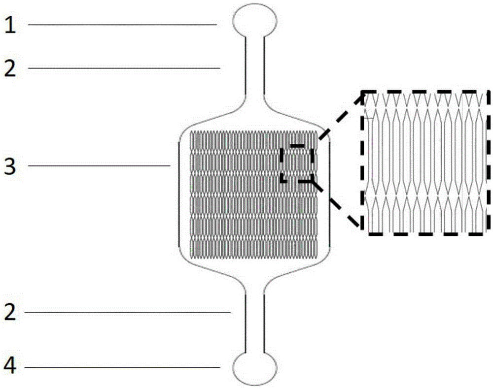
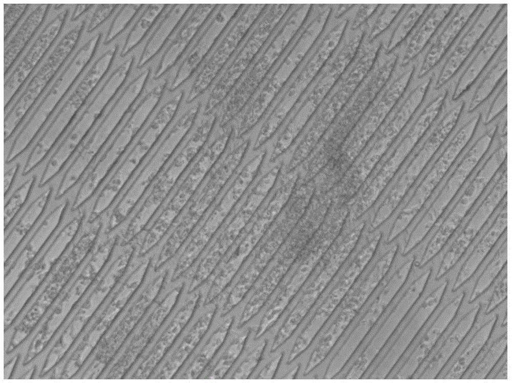
[0040] Using the above microfluidic chip, the structure is as follows figure 1 shown. Prepare Matrigel working solution with a concentration of 150 μg / mL in DMEM-F12 medium, inject it into the chip through the cell inlet pool, let it stand at 4°C overnight, and remove the working solution after 24 hours. After hiPSC-CM cells were digested, diluted to a concentration of 5×10 6 cells / mL cell suspension, add 100 μL of cell suspension into the chip through the cell inlet pool, under the modification of Matrigel, the cells quickly adhere to the wall and evenly spread on the bottom of the cell culture chamber ( figure 2 ), when the uniform distribution of cells in the cell culture chamber was observed under an optical microscope, the chip was immediately moved into a carbon dioxide incubator to continue culturing. After the hiPSC-CM cells were completely attached to the bottom of the cell culture chamber, the cell...

Embodiment 2

[0042] Characterization of functional proteins in human heart model cells

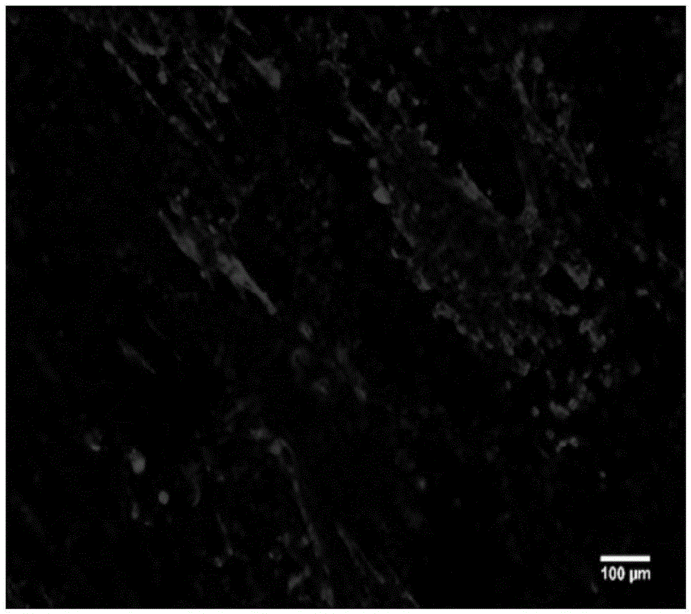
[0043] Using the microfluidic chip designed and manufactured by the laboratory, the structure is as follows: figure 1shown. After chip modification, the same cell inoculation and culture methods as in Example 1 were used to establish a heart model. After 7 days of perfusion, immunofluorescence staining was performed on the cells, and the detected protein was troponin (cTnT). The method is as follows: cells were fixed with 4% paraformaldehyde, washed three times with PBS buffer, 10 min each time; treated with 0.1% triton X-100 porogen for 10 min, washed three times with PBS buffer, 10 min each time; blocked with goat serum for 1 h, The primary antibody (mouse anti-human cTnT) was diluted 1:100, incubated overnight at 4°C, washed three times with PBS buffer for 10 minutes each time; the secondary antibody (AlexaFluor488-labeled goat anti-mouse IgG) was diluted 1:100, incubated at room temperature and p...


PUM


Abstract

The invention provides a method for establishing a micro-fluidic chip based human heart model. A micro-fluidic chip mainly comprises a cell inlet pool, channels, a cell culture chamber and an outlet pool, wherein the cell culture chamber is connected to the cell inlet pool and the cell outlet pool through straight channels; and the cell culture chamber has a structure having repeated hexagonal bulges. The method for establishing a human heart model comprises the following steps: (1) directionally differentiating hiPSC into myocardial cells; (2) modifying the chip; (3) inoculating and culturing cells in the chip; and (4) establishing the human heart model. The model is used for observing representations of cell activity investigation and function changes in the chip, and can be used for human heart auxology researches and drug cardiotoxicity. The micro-fluidic chip based human heart model is used for observing morphologic changes and arrangement of myocardial cells having a micro-structural surface under the perfusion condition at different flow-speeds, especially for the influence of blood flow shearing force and matrix surface microstructure on the myocardial cell developmental maturity in the heart developing process.

Description

technical field [0001] The invention belongs to the technical field of applying microfluidic chip technology to cell biology research of tissue bionics and real-time monitoring, and specifically relates to a method for establishing a human heart model based on a microfluidic chip. Background technique [0002] Cardiovascular disease is the number one cause of death in the world, killing more people each year than any other disease. During drug development, more than one-third of drugs fail safety testing due to cardiotoxicity and are withdrawn. A large number of animal experiments are required before clinical trials of drugs. These experiments are generally very expensive, time-consuming, and ethically controversial. Moreover, due to species differences, they cannot accurately reflect the effects of drugs in the human body, which often leads to the failure of drugs in clinical trials. . Because the traditional two-dimensional cell culture model cannot simulate the microenv...

Claims


Application Information

Patent Timeline
IPC IPC(8): C12M3/00C12N5/077C12Q1/02C12Q1/68
CPCC12M23/16C12M25/06C12N5/0657C12N2506/03C12Q1/6883C12Q2600/136C12Q2600/158G01N33/5023G01N33/5026G01N33/5061
Inventor 秦建华徐聪于跃王丽
Owner DALIAN INST OF CHEM PHYSICS CHINESE ACAD OF SCI
Why Eureka
Try Eureka