Plaque Stability Measurement System Based on Optical Coherence Tomography

A stability and plaque technology, applied in the field of fast, efficient and accurate measurement systems for plaque stability, can solve problems such as guide wire failure, non-uniform rotation distortion, cost increase, etc., to achieve comprehensive and efficient measurement analysis, improve Accuracy, time saving effect

Active Publication Date: 2021-08-24
PULSE MEDICAL IMAGING TECH (SHANGHAI) CO LTD
View PDF6 Cites 0 Cited by
  • Summary
  • Abstract
  • Description
  • Claims
  • Application Information

AI Technical Summary

Problems solved by technology

[0008] However, the above-mentioned equipment and method also have several limitations: 1) the length of the hard front end of the guide wire is long, which may cause the guide wire to fail to pass through, and easily cause nonuniform rotational distortion (Nonuniform Rotational Distortion) in the image; 2) the above-mentioned system The imaging frame rate is limited, which is mainly due to the limitations of the existing ultrasonic pulse generator and the repetition rate of the scanning laser light source; 3) The combination of the two imaging systems is bound to increase the cost, which is not conducive to the popularization of this method
[0009] Furthermore, there is currently no method and system for analyzing plaque stability by obtaining plaque lipid core size and plaque stress information only through optical coherence tomography

Method used


Image

  • Plaque Stability Measurement System Based on Optical Coherence Tomography
  • Plaque Stability Measurement System Based on Optical Coherence Tomography
  • Plaque Stability Measurement System Based on Optical Coherence Tomography

Examples


Embodiment 1

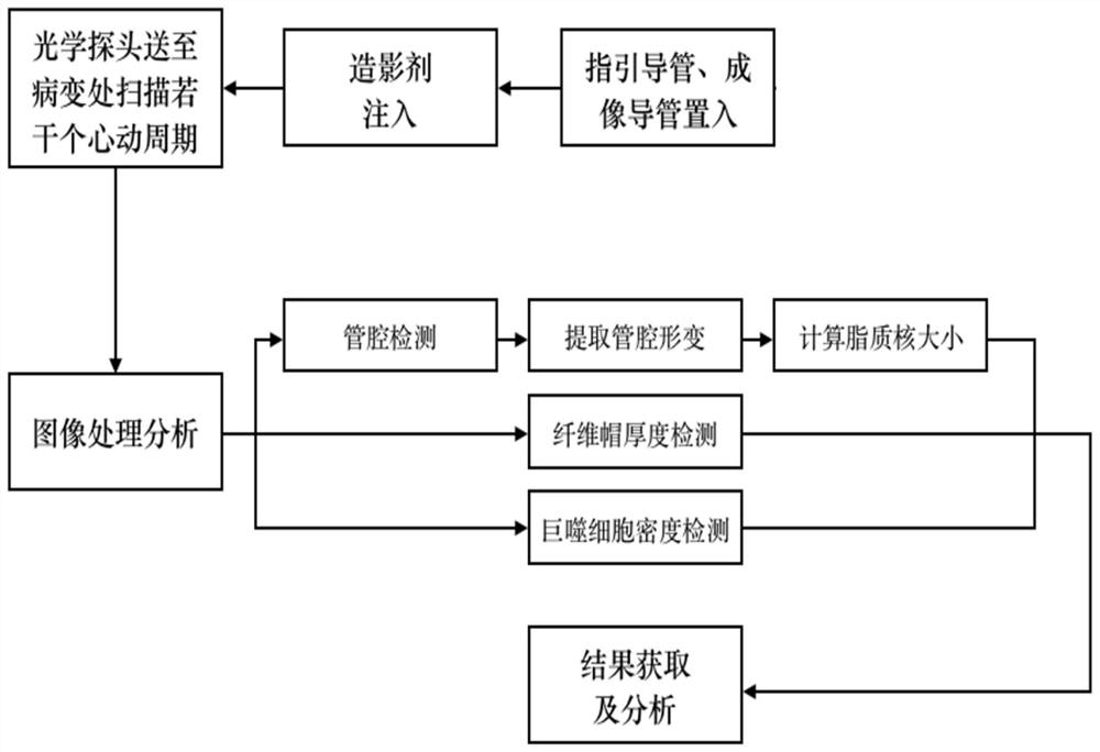
[0046] In order to further understand the present invention, the present invention will be further elaborated and illustrated below in conjunction with the accompanying drawings and embodiments. figure 1 It is a flow chart of the method of the present invention. In a specific embodiment, the method can be summarized as follows: the optical probe is sent to the lesion to scan several cardiac cycles, and the deformation of the vessel lumen at the same site at multiple key time points of the cardiac cycle is obtained. ; It is preferred to obtain the lipid core size and plaque stress information of the plaque site through the table look-up method; combined with the measurement and analysis of the plaque fibrous cap thickness, macrophage infiltration degree, etc. by traditional optical coherence tomography, the stability of the plaque is analyzed. A more complete and comprehensive analysis.

[0047] Table 1 is an example of the form used by the above table look-up method. The maxim...

Embodiment 2

[0078] In another specific embodiment, the plaque stability on a specific blood vessel is analyzed by using the method of the present invention. Specifically, this method can be implemented through the following steps:

[0079] (1), select the left circumflex branch of the heart as the vessel of interest;

[0080] (2), determine the position of the plaque in the left circumflex branch;

[0081] (3) During the process of optical coherence tomography, the scanning device stays at the position determined in step (2), and scans multiple cardiac cycles, so as to obtain tomographic images where the plaque is located at multiple moments in the cardiac cycle;

[0082] (4), after the scanning device stays at the position of the plaque determined in step (2) for multiple cardiac cycles, the traditional optical coherence tomography process is continued;

[0083] (5) Obtain multi-moment tomographic imaging images within the time period of step (3), select N time points in the cardiac cy...

Embodiment 3

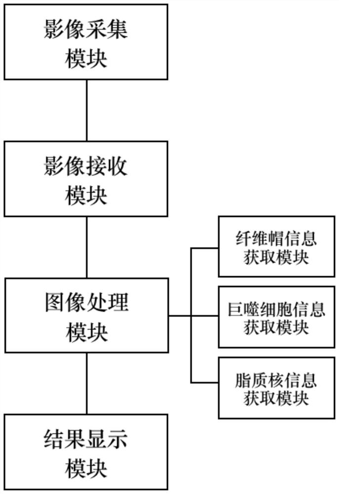
[0088] In yet another specific embodiment, the present invention also provides a plaque stability measurement system based on optical coherence tomography, the system comprising:

[0089] The image acquisition module is used for coronary artery image acquisition and generates image signals;

[0090] The image receiving module is used to receive the image signal generated by the image acquisition module and transmit it to the image processing module;

[0091] The image processing module processes and analyzes the received image signal. It includes the measurement and analysis of visible features and latent features of plaques. The measurement and analysis of visible features of plaques includes the acquisition of fibrous cap thickness and macrophage density. Lipid core size and plaque stress;

[0092] The result display module is used to display the measurement and analysis results of the image processing module.

[0093] Preferably, the image processing module performs visi...


PUM


Abstract

The present invention provides a plaque stability measurement method and system based on optical coherence tomography. The system includes: an image acquisition module, an image receiving module, an image processing module, and a result display module, wherein: the image acquisition module is used for coronary Image acquisition and image signal generation; the image receiving module is used to receive the image signal generated by the image acquisition module and transmit it to the image processing module; the image processing module is used to perform image processing and analysis on the received image signal, including fiber cap information acquisition module, macrophage information acquisition module and lipid nucleus information acquisition several sub-modules; the result display module is used to display the measurement and analysis results of the image processing module. The invention breaks through the limitation that the traditional optical coherence tomography cannot obtain lipid nucleus size parameters, and adds information on lipid nucleus size and plaque stress while ensuring the high-resolution results unique to optical coherence tomography.

Description

technical field [0001] The invention is applied in the field of medical equipment and signal processing, and in particular relates to an image processing and analysis system based on optical coherence tomography, and a fast, efficient and accurate measurement system for plaque stability based on optical coherence tomography images and method. Background technique [0002] Optical coherence tomography (OCT) is an emerging high-resolution tomography technique. Similar to the principle of intravascular ultrasound (Intravascular Ultrasound, IVUS), this technology uses near-infrared rays as the light source, uses the interference law of light waves to perform imaging, and converts the optical signal of the beam scanning the tissue into an electrical signal, which is displayed as a gray image or a pseudo image after computer processing. 2D and 3D images of colormaps. Because the wavelength of light is shorter than that of sound waves, optical coherence tomography has higher reso...

Claims


Application Information

Patent Timeline
Patent Type & Authority Patents(China)
IPC IPC(8): A61B5/00A61B5/02
CPCA61B5/0033A61B5/0066A61B5/02007A61B5/7235
Inventor 黄佳悦涂圣贤田峰余炜张素
Owner PULSE MEDICAL IMAGING TECH (SHANGHAI) CO LTD
Why Eureka
Try Eureka